Copyright
©The Author(s) 2020.
World J Gastrointest Oncol. Oct 15, 2020; 12(10): 1177-1194
Published online Oct 15, 2020. doi: 10.4251/wjgo.v12.i10.1177
Published online Oct 15, 2020. doi: 10.4251/wjgo.v12.i10.1177
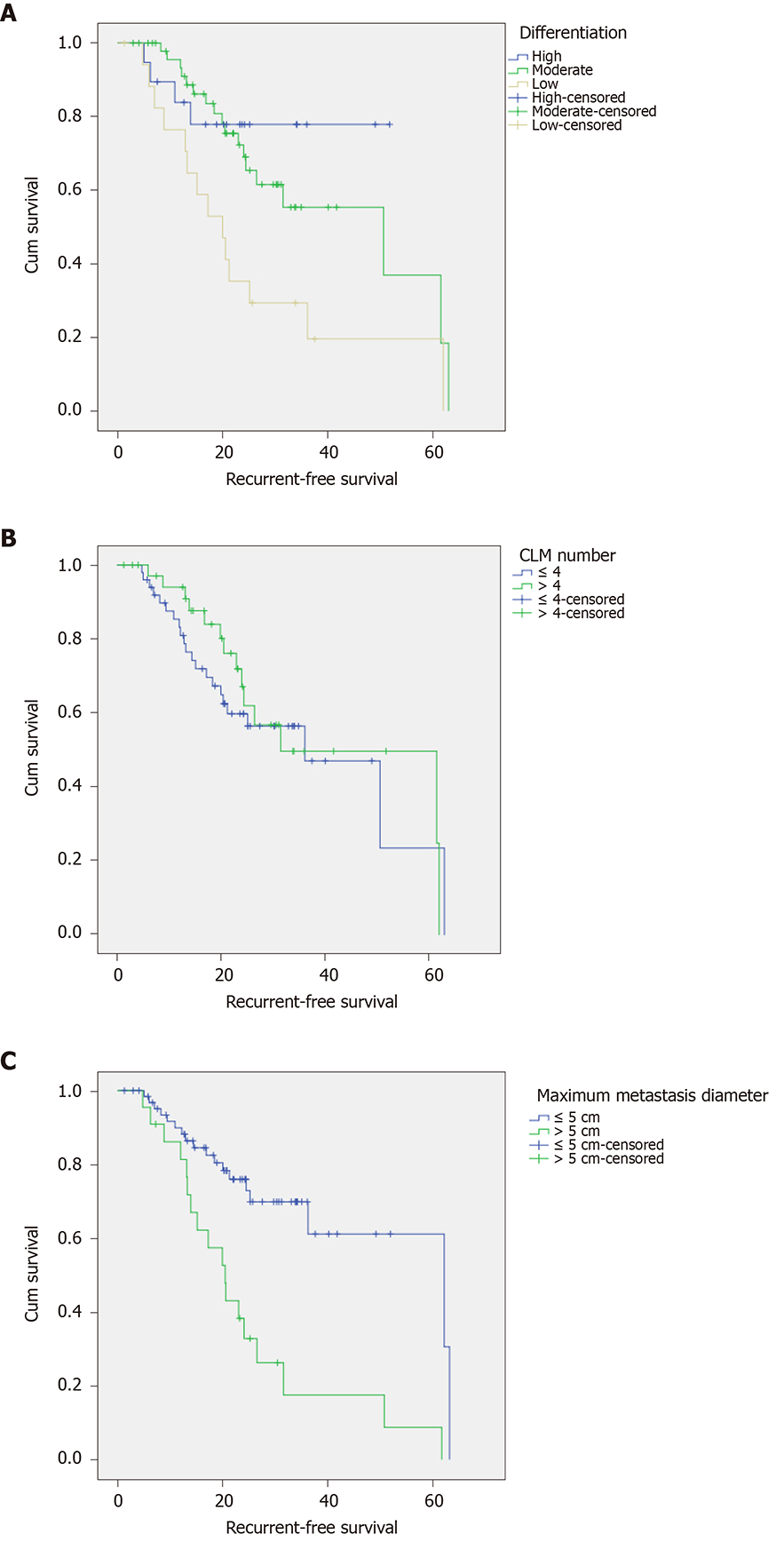
Figure 1 Survival curves of patients.
A: Different differentiation degrees of the primary tumor; B: Colorectal liver metastases ≤ 4 and> 4; C: Colorectal liver metastases with a maximum metastasis diameter of ≤ 5 cm and > 5 cm. CLM: Colorectal liver metastases.
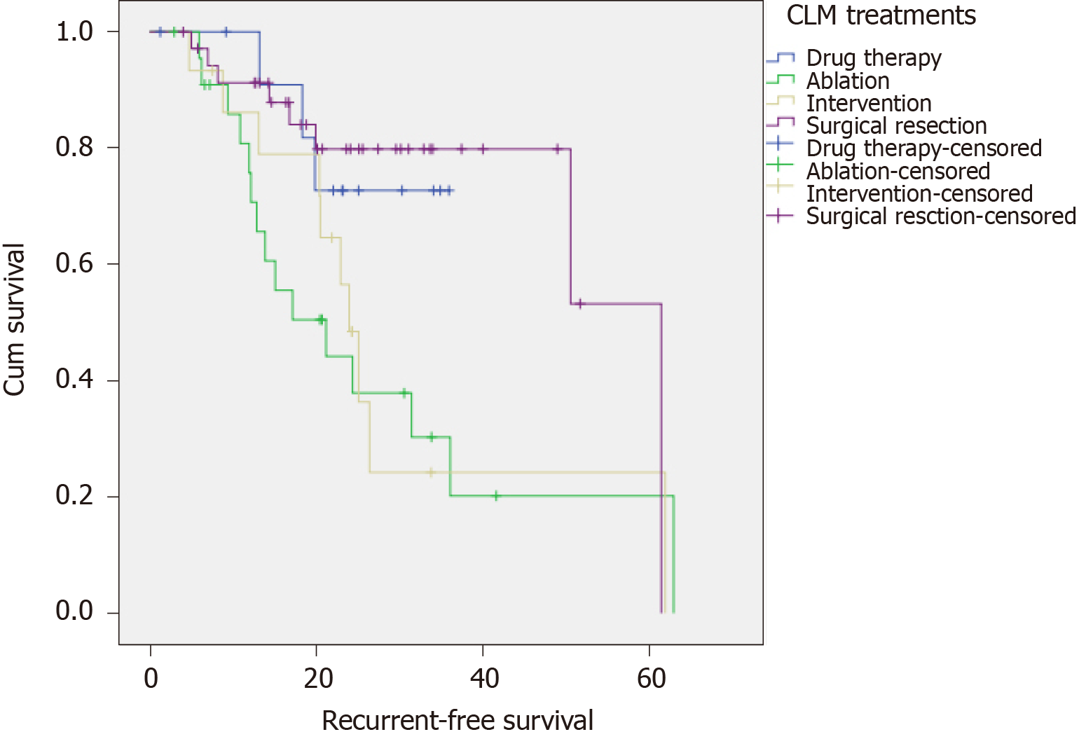
Figure 2 Survival curves for different treatments.
CLM: Colorectal liver metastases.
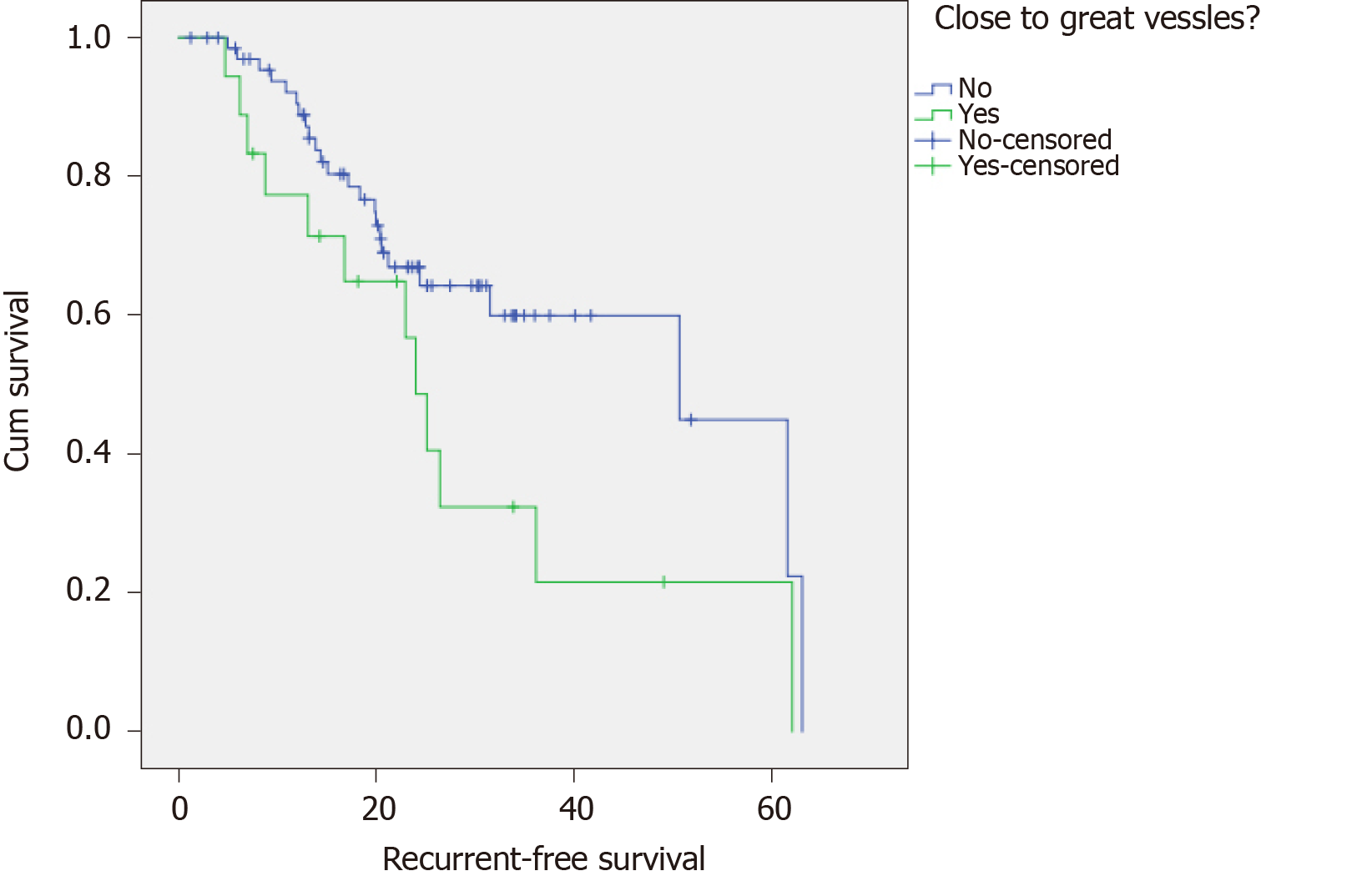
Figure 3 Survival curves of colorectal liver metastases near or not near great vessels.
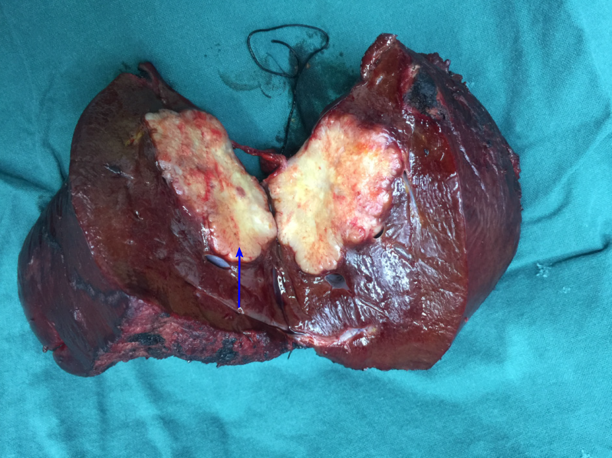
Figure 4 Surgical resection of colorectal liver metastases.
The arrow indicates metastases.
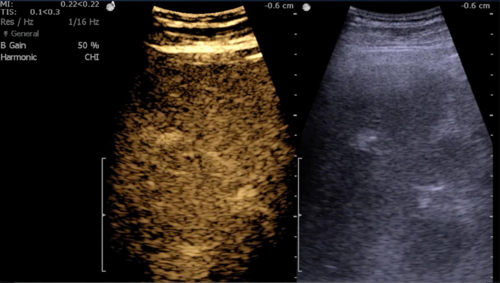
Figure 5 Intraoperative ultrasound positioning.
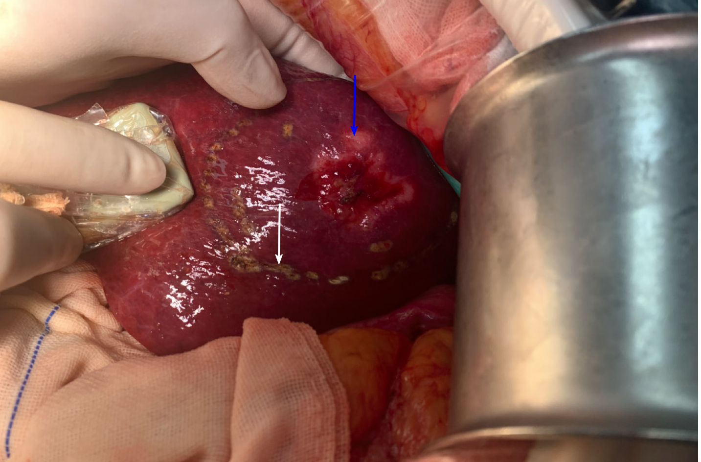
Figure 6 Intraoperative ultrasound localization.
Blue arrow represents colorectal liver metastases, and white arrow represents electrosurgical markers after ultrasound localization.
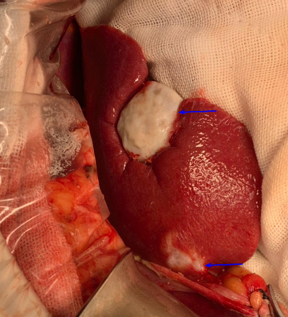
Figure 7 Radiofrequency ablation.
Arrows indicate necrosis of colorectal liver metastases.
- Citation: Ma ZH, Wang YP, Zheng WH, Ma J, Bai X, Zhang Y, Wang YH, Chi D, Fu XB, Hua XD. Prognostic factors and therapeutic effects of different treatment modalities for colorectal cancer liver metastases. World J Gastrointest Oncol 2020; 12(10): 1177-1194
- URL: https://www.wjgnet.com/1948-5204/full/v12/i10/1177.htm
- DOI: https://dx.doi.org/10.4251/wjgo.v12.i10.1177


