Copyright
©The Author(s) 2019.
World J Gastrointest Oncol. Oct 15, 2019; 11(10): 830-841
Published online Oct 15, 2019. doi: 10.4251/wjgo.v11.i10.830
Published online Oct 15, 2019. doi: 10.4251/wjgo.v11.i10.830
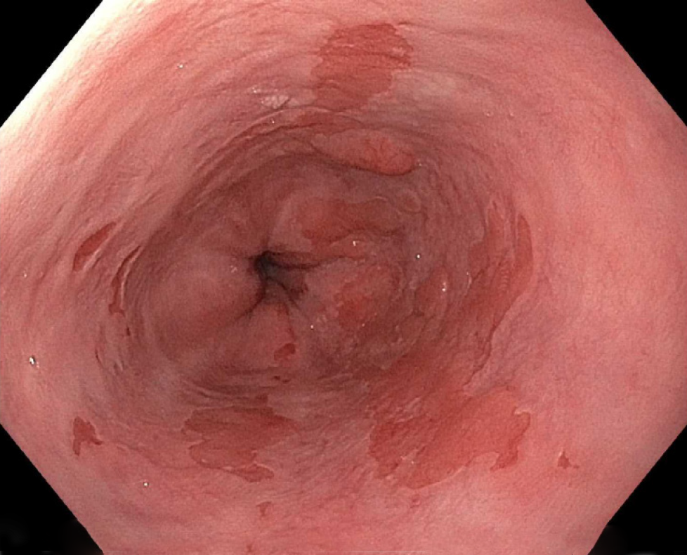
Figure 1 Barrett’s esophagus with nodularity.
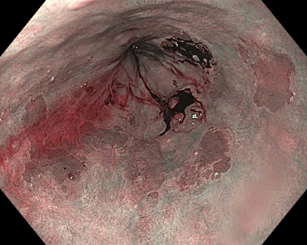
Figure 2 Narrow-band imaging of Barrett’s esophagus.
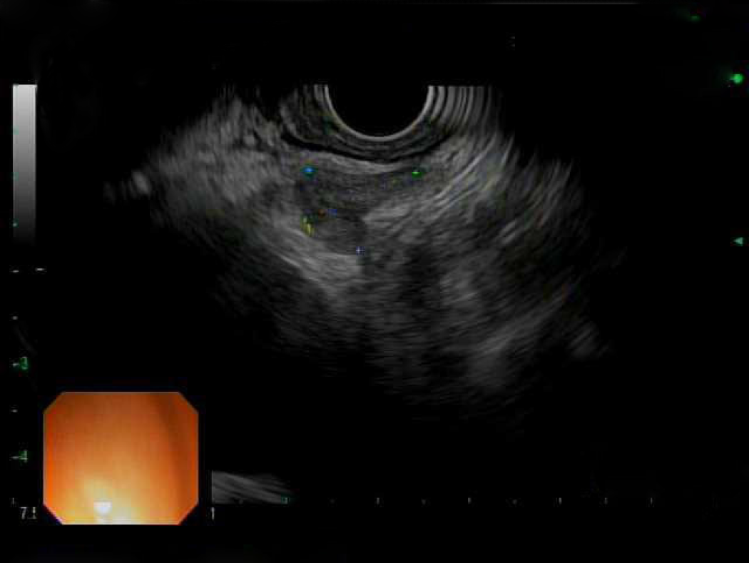
Figure 3 Endoscopic ultrasound image of subcarinal lymph node.
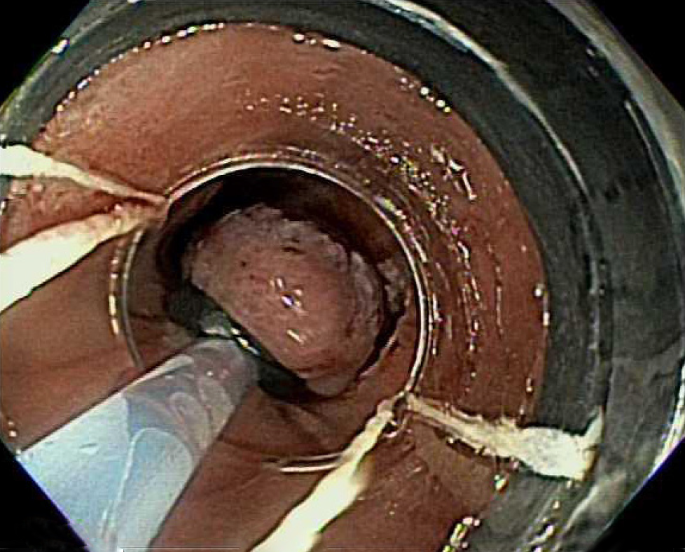
Figure 4 Band-ligation method of endoscopic mucosal resection.
Figure 5 Post-endoscopic mucosal resection.
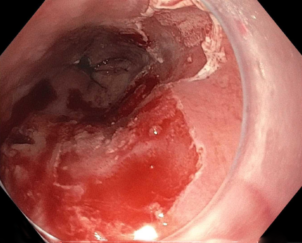
Figure 6 Post-radiofrequency ablation.
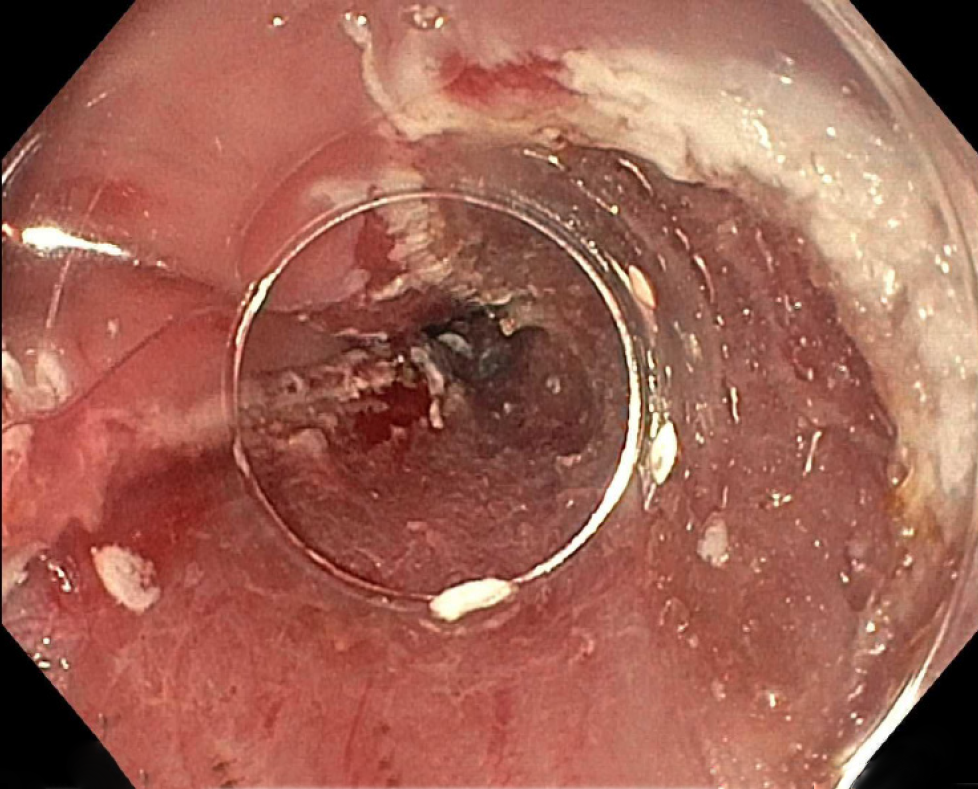
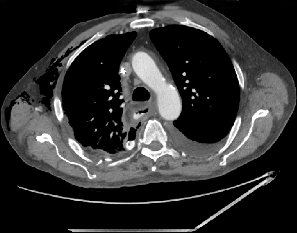
Figure 7 Post-operative anastomotic leak.
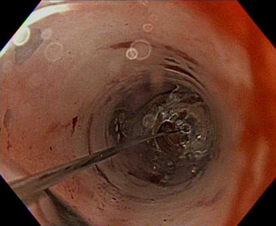
Figure 8 Esophageal balloon dilation.
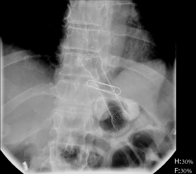
Figure 9 Self-expanding metal stent esophageal stent.
- Citation: Ahmed O, Ajani JA, Lee JH. Endoscopic management of esophageal cancer. World J Gastrointest Oncol 2019; 11(10): 830-841
- URL: https://www.wjgnet.com/1948-5204/full/v11/i10/830.htm
- DOI: https://dx.doi.org/10.4251/wjgo.v11.i10.830


