Copyright
©The Author(s) 2016.
World J Gastrointest Endosc. Dec 16, 2016; 8(20): 741-755
Published online Dec 16, 2016. doi: 10.4253/wjge.v8.i20.741
Published online Dec 16, 2016. doi: 10.4253/wjge.v8.i20.741
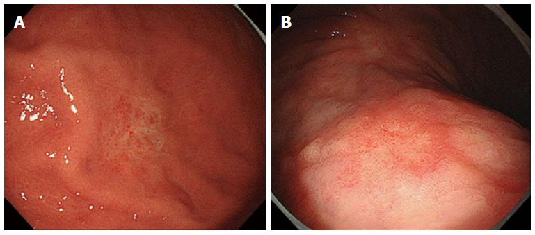
Figure 1 High definition white light endoscopy view.
A: A depressed lesion with mucosal discoloration due to early gastric cancer; B: High definition white light endoscopy view of early gastric cancer.
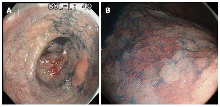
Figure 2 Mucosal irregularities and boundaries of a lesion.
A: Gastric adenoma accentuated by indigo carmine; B: Early gastric cancer accentuated by indigo carmine.
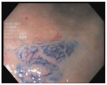
Figure 3 Gastric intestinal metaplasia highlighted by methylene blue.
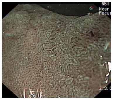
Figure 4 Gastric intestinal metaplasia highlighted by narrow band imaging using the EXERA III system with dual focus.
Figure 5 Best visualized with optical magnification.
A: Magnifying narrow band image of gastric intestinal metaplasia showing, the light blue crest sign, surrounding central area of early gastric cancer; B: Magnifying narrow band image of early gastric cancer; C: Magnifying narrow band image of early gastric cancer.
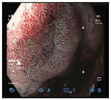
Figure 6 Image of early gastric cancer visualized using narrow band imaging with digital magnification and dual focus imaging.
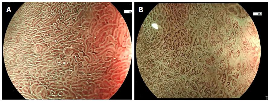
Figure 7 Performance characteristics.
A: Image of gastric intestinal metaplasia visualized by blue laser imaging with optical magnification; B: Image of early gastric cancer visualized by blue laser imaging with optical magnification.
- Citation: Hussain I, Ang TL. Evidence based review of the impact of image enhanced endoscopy in the diagnosis of gastric disorders. World J Gastrointest Endosc 2016; 8(20): 741-755
- URL: https://www.wjgnet.com/1948-5190/full/v8/i20/741.htm
- DOI: https://dx.doi.org/10.4253/wjge.v8.i20.741


