Copyright
©The Author(s) 2019.
World J Gastrointest Endosc. Aug 16, 2019; 11(8): 454-471
Published online Aug 16, 2019. doi: 10.4253/wjge.v11.i8.454
Published online Aug 16, 2019. doi: 10.4253/wjge.v11.i8.454
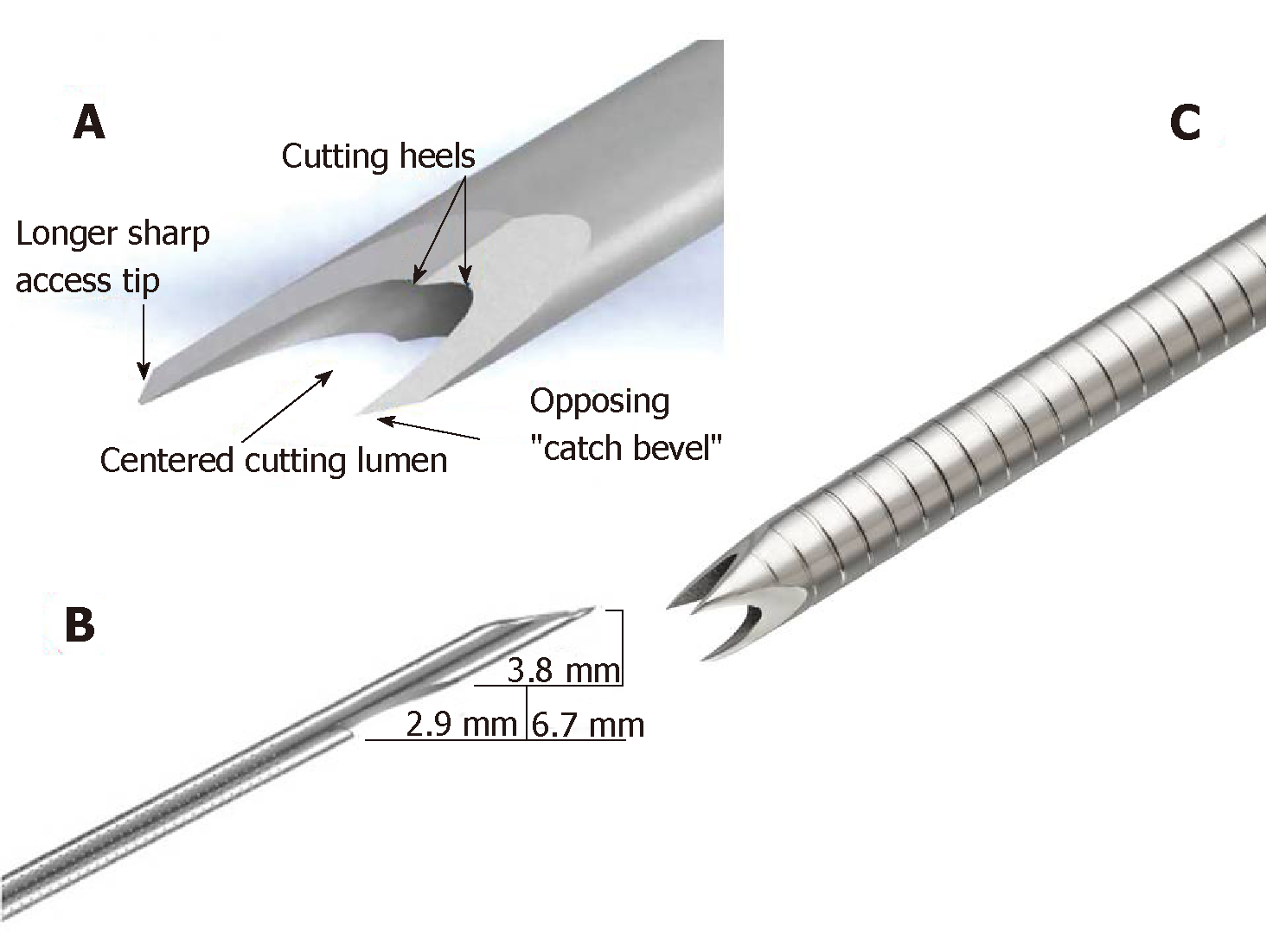
Figure 1 Fine needle biopsy needles types.
A: Acquire (Boston Scientific, Marlborough, MA, United States) needle; B: SharkCore™ (Medtronic Inc., Sunnyvale, CA, United States) needle; C: ProCore™ (Wilson-Cook Medical Inc., Winston-Salem, NC, United States) needle.
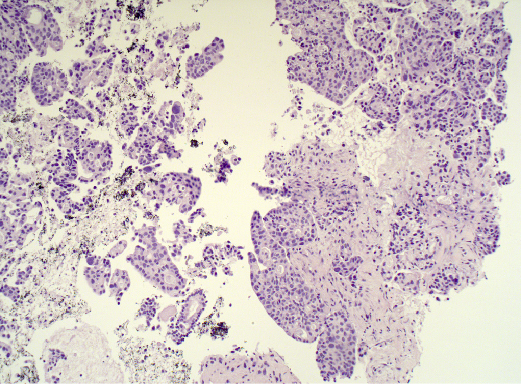
Figure 2 Fine needle biopsy sample of pancreatic adenocarcinoma, which clearly shows the preserved histological architecture of the malignant tissue (hematoxylin and eosin staining, 10 ×).
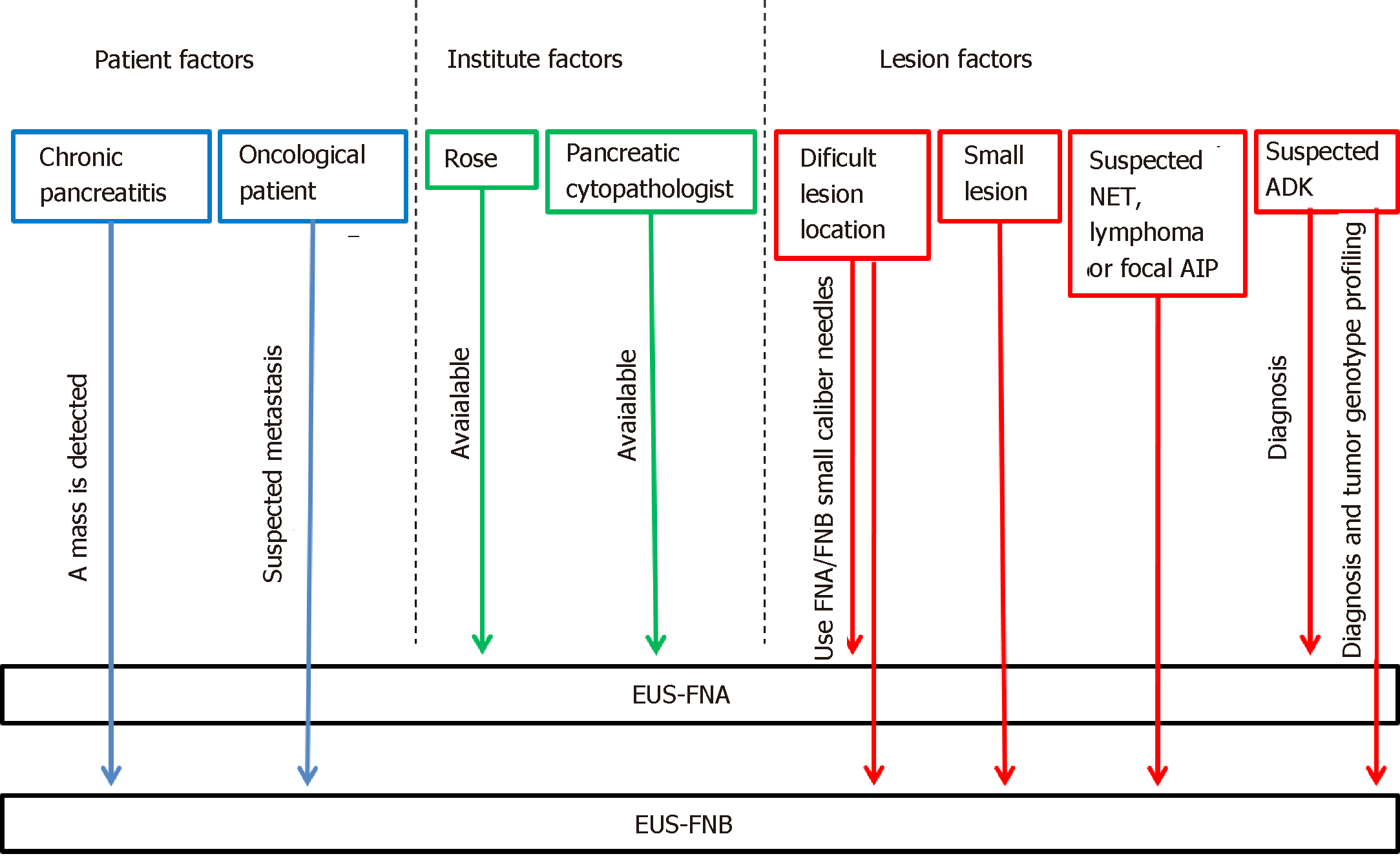
Figure 3 A practical flow chart for selecting among the available needles in each scenario (pancreatic neuroendocrine tumors, pancreatic neuroendocrine tumors; autoimmune pancreatitis, autoimmune pancreatitis; adenocarcinoma, adenocarcinoma).
ROSE: Rapid on site evaluation; EUS-FNA: Endoscopic ultrasound guided-fine needle aspiration; EUS-FNB: Endoscopic ultrasound guided-fine needle biopsy; ADK: Adenocarcinoma; AIP: Autoimmune pancreatitis.
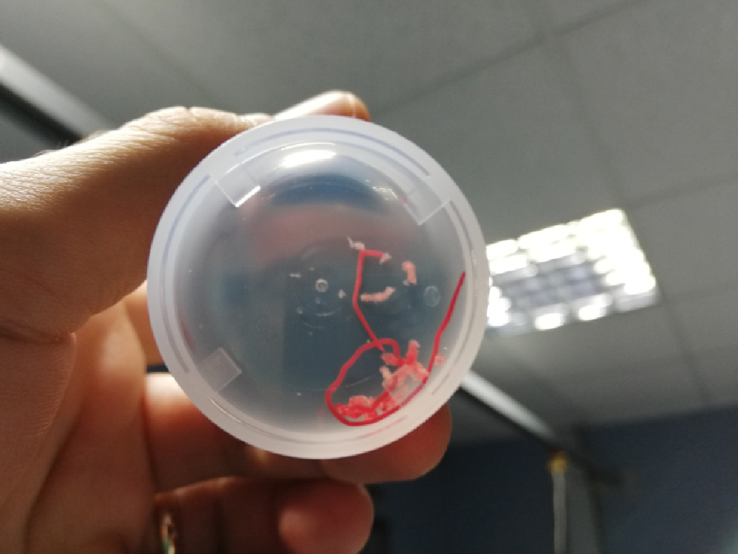
Figure 4 Endoscopic ultrasound guided-fine needle biopsy sample of a pancreatic lesion, obtained by using ProCore 22 G needle.
- Citation: Conti CB, Cereatti F, Grassia R. Endoscopic ultrasound-guided sampling of solid pancreatic masses: the fine needle aspiration or fine needle biopsy dilemma. Is the best needle yet to come? World J Gastrointest Endosc 2019; 11(8): 454-471
- URL: https://www.wjgnet.com/1948-5190/full/v11/i8/454.htm
- DOI: https://dx.doi.org/10.4253/wjge.v11.i8.454


