Copyright
©The Author(s) 2016.
World J Stem Cells. Mar 26, 2016; 8(3): 106-117
Published online Mar 26, 2016. doi: 10.4252/wjsc.v8.i3.106
Published online Mar 26, 2016. doi: 10.4252/wjsc.v8.i3.106
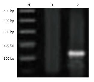
Figure 1 An agarose gel electrophoresis of DNA fragments showed SRY gene in recipient female rats for bone marrow derived mesenchymal stem cells in Parkinson’s disease model.
Lane (M) represents DNA ladder; Lane (1) represents ovariectomized control sample; Lane (2) represents sample from PD group treated with BM-MSCs. PD: Parkinson’s disease; BM-MSCs: Bone marrow derived mesenchymal stem cells.
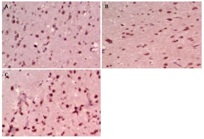
Figure 2 Immunohistochemical examination of survivin expression in Parkinson’s disease model groups.
A: Ovariectomized control; B: PD untreated; C: PD + BM-MSCs. PD: Parkinson’s disease; BM-MSCs: Bone marrow derived mesenchymal stem cells.
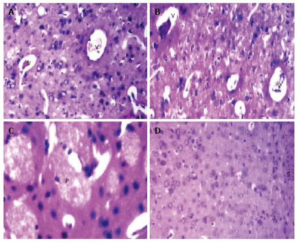
Figure 3 Photomicrograph of brain section of: A: Ovariectomized control group shows congestion in blood vessels of striatum (v) (H and E × 80); B: untreated Parkinson’s disease (PD) group shows congestion in blood vessels and capillaries of striatum (v) (H and E × 80); C: Untreated PD: Parkinson’s disease group shows hyalinization with plaques formation in the matrix of striatum (H and E × 160); and D: PD group treated with bone marrow derived mesenchymal stem cells shows intact histological structure of the striatum (H and E × 80).
- Citation: Ahmed HH, Salem AM, Atta HM, Eskandar EF, Farrag ARH, Ghazy MA, Salem NA, Aglan HA. Updates in the pathophysiological mechanisms of Parkinson’s disease: Emerging role of bone marrow mesenchymal stem cells. World J Stem Cells 2016; 8(3): 106-117
- URL: https://www.wjgnet.com/1948-0210/full/v8/i3/106.htm
- DOI: https://dx.doi.org/10.4252/wjsc.v8.i3.106


