Copyright
©2014 Baishideng Publishing Group Co.
World J Stem Cells. Jan 26, 2014; 6(1): 33-42
Published online Jan 26, 2014. doi: 10.4252/wjsc.v6.i1.33
Published online Jan 26, 2014. doi: 10.4252/wjsc.v6.i1.33
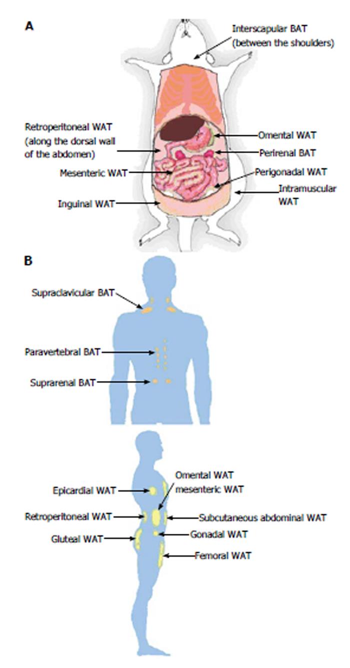
Figure 1 Locations of adipose tissue depots in a mouse (A) and an adult human (B).
A: Subcutaneous (inguinal and intramuscular), visceral (mesenteric, omental, perigonadal and retroperitoneal) and brown (interscapular and perirenal) adipose tissue depots are shown in a mouse model; B: Subcutaneous (abdominal, femoral and gluteal), visceral (epicardial, gonadal, mesenteric, omental and retroperitoneal) and brown (paravertebral, supraclavicular and suprarenal) adipose tissue depots are shown in a human model. WAT: White adipose tissue; BAT: Brown adipose tissue.
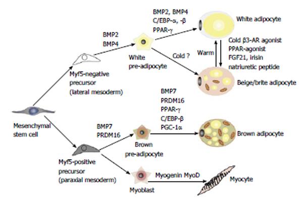
Figure 2 Differentiation into white, beige or brown adipocytes.
Previously, white and brown adipocytes were thought to be derived from the same precursor cell. However, recent studies demonstrated that brown fat shares a progenitor cell (Myf5+) with skeletal muscle and not with white adipocytes. The Myf5+ precursors are induced to transform into mature brown adipocytes by bone morphogenetic protein 7 (BMP7), peroxisome proliferator-activated receptor-γ (PPAR-γ) and CCAAT/enhancer-binding proteins (C/EBPs) in cooperation with the transcriptional co-regulator PR domain-containing 16 (PRDM16) and PGC-1α. White adipocytes can also be transformed to brown-like adipocytes, called beige/brite adipocytes, by cold exposure, a β-adrenergic agonist or a PPAR-γ agonist. AR: adrenergic receptor; FGF21: Fibroblast growth factor 21; PGC-1α: Peroxisome proliferator activated receptor gamma coactivator 1 alpha.
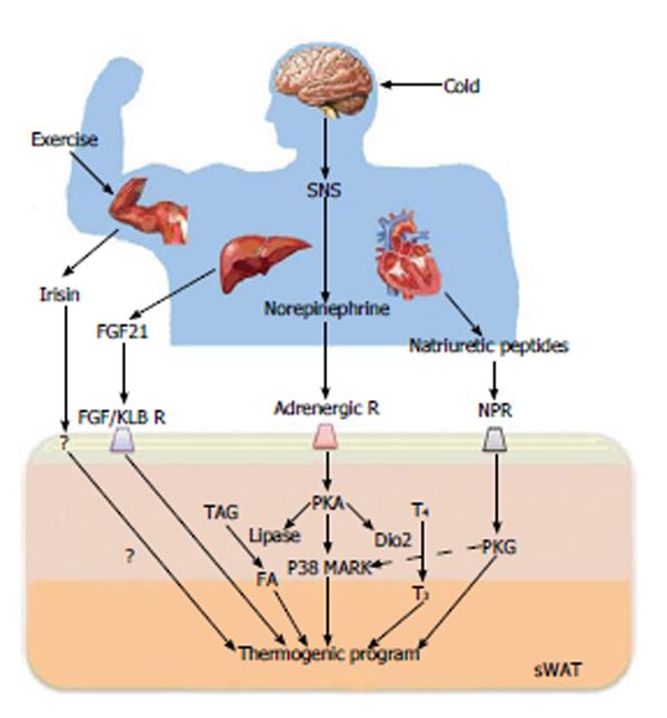
Figure 3 Key regulators of the browning process and their action mechanisms.
Browning is induced by sympathetic nervous system (SNS)-independent or SNS-dependent signals. These signals sometimes synergistically or competitively influence the activation of browning of subcutaneous white adipose tissue (sWAT). Irisin is a newly discovered myokine and is released by skeletal muscle during exercise. Irisin induces the browning process of sWAT. Fibroblast growth factor-21 (FGF21), a hormonal factor from the liver, directly activates the thermogenic process via interaction with the FGF receptor/β-Klotho (KLB) complex. The norepinephrine secreted by the SNS in response to thermogenic stimuli induces the activation of adrenergic receptor(s). The adrenergic receptor-mediated signal increases the level of intracellular cAMP and activates cAMP-dependent protein kinase A (PKA). Subsequently, PKA activates p38 MAP kinase (p38 MAPK) and 5’-deiodinase 2 (Dio2), which catalyzes the conversion of thyroxine (T4) into the active form 3,5,3’-tri-iodothyronine (T3). Then, it ultimately induces the gene process for thermogenic activation. Natriuretic peptides (NPs) originating from the heart activate the thermogenic process through binding to the NP receptor, activation of protein kinase G (PKG) and the subsequent activation of p38 MAPK and NPR. NPR: Natriuretic peptides receptor; TAG: Triacylglycerol; FA: Fatty acid.
- Citation: Park A, Kim WK, Bae KH. Distinction of white, beige and brown adipocytes derived from mesenchymal stem cells. World J Stem Cells 2014; 6(1): 33-42
- URL: https://www.wjgnet.com/1948-0210/full/v6/i1/33.htm
- DOI: https://dx.doi.org/10.4252/wjsc.v6.i1.33


