Copyright
©The Author(s) 2024.
World J Stem Cells. Feb 26, 2024; 16(2): 191-206
Published online Feb 26, 2024. doi: 10.4252/wjsc.v16.i2.191
Published online Feb 26, 2024. doi: 10.4252/wjsc.v16.i2.191
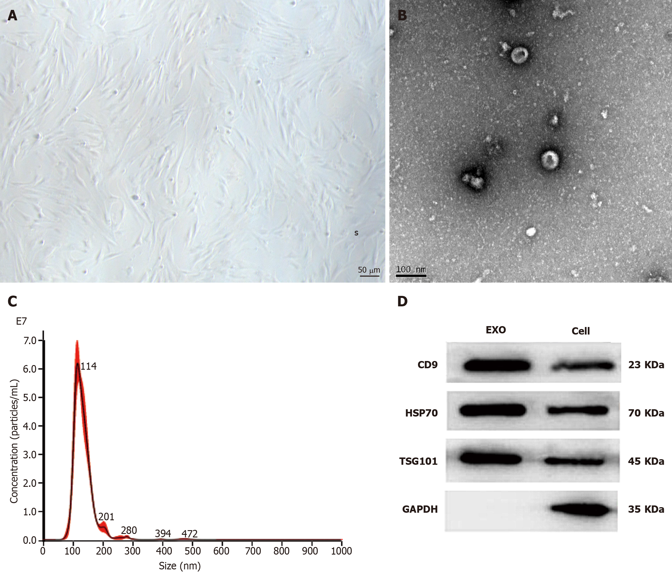
Figure 1 Morphological observation of bone marrow-derived mesenchymal stem cells and identification of bone marrow-derived mesenchymal stem cells-exosomes.
A: The bone marrow-derived mesenchymal stem cells (BMSCs) had a homogeneous spindle morphology; B: The BMSC-exosomes (Exos) were approximately 60–160 nm in diameter and had a sub-circular, typical teatoid appearance; C: The nanoparticle tracking analysis results showed that the concentration of BMSC-Exos was 3.43 × 10⁹/mL and their average particle size was 131.9 nm; D: The results of the western blotting assays showed that the expression of CD9, heat shock protein 70, and tumor necrosis factor-alpha-stimulated gene 101 was positive. Exo: Exosomes; HSP: Heat shock protein; TSG: Tumor necrosis factor-alpha-stimulated gene.
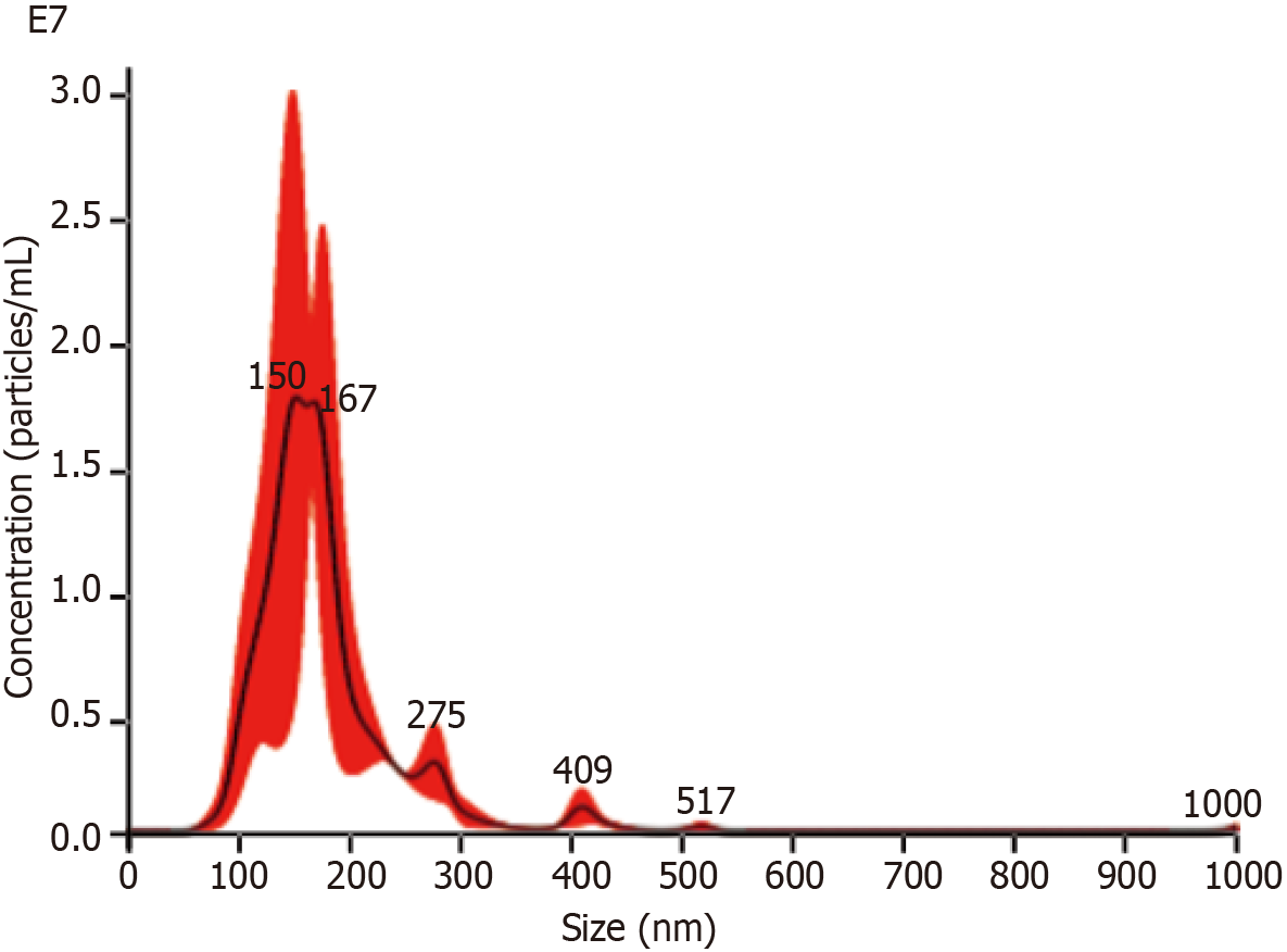
Figure 2
The results of nanoparticle tracking analysis of bone marrow-derived mesenchymal stem cells-miR-29a-Exos.
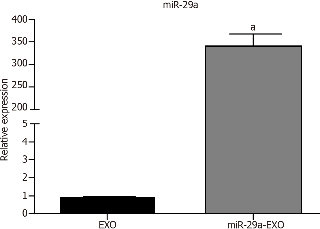
Figure 3 The results of the quantitative reverse transcription polymerase chain reaction assay.
aP < 0.001. Exo: Exosomes.
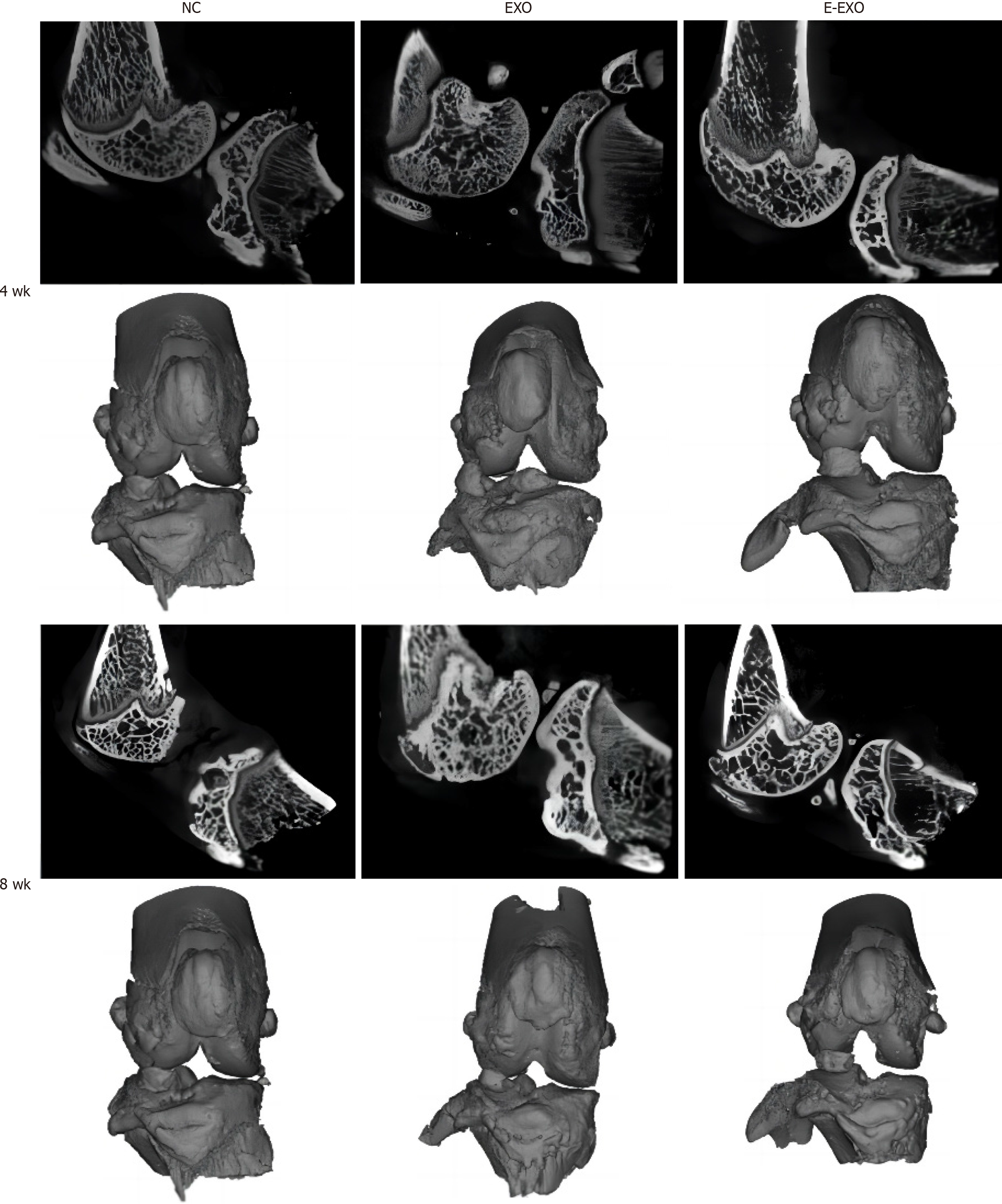
Figure 4 Micro-computed tomography and three-dimensional reconstruction of the knee joint of rats.
Micro-computed tomography images of the knee joint and the 3D reconstructed images of the corresponding specimens for each group of rats at four weeks and eight weeks are presented. Exo: Exosomes; NC: Negative control.
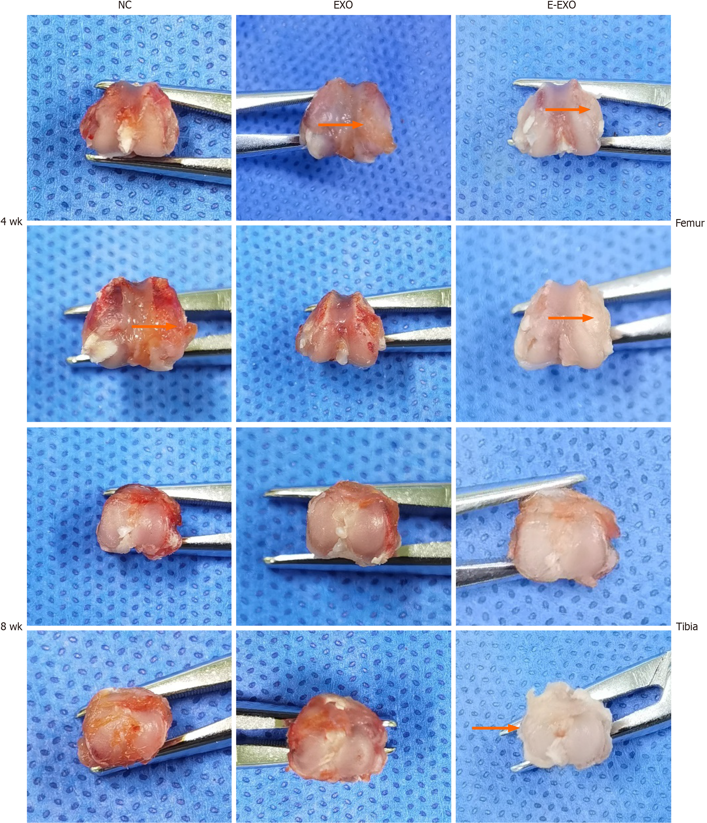
Figure 5 Photographs of gross specimens of rat knee joints.
Exo: Exosomes; NC: Negative control.
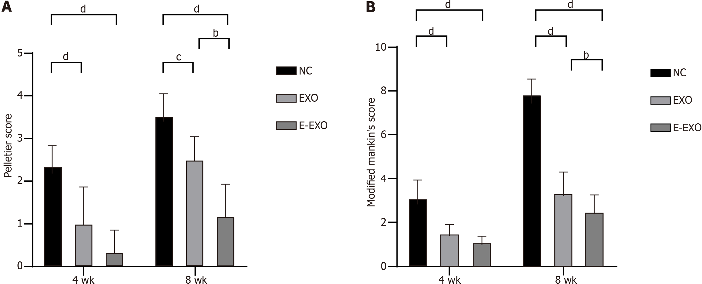
Figure 6 Pelletier gross scores and the modified Mankin score.
A: Pelletier gross scores; B: The modified Mankin score. bP < 0.01, cP < 0.05, dP < 0.01. Exo: Exosomes; NC: Negative control.
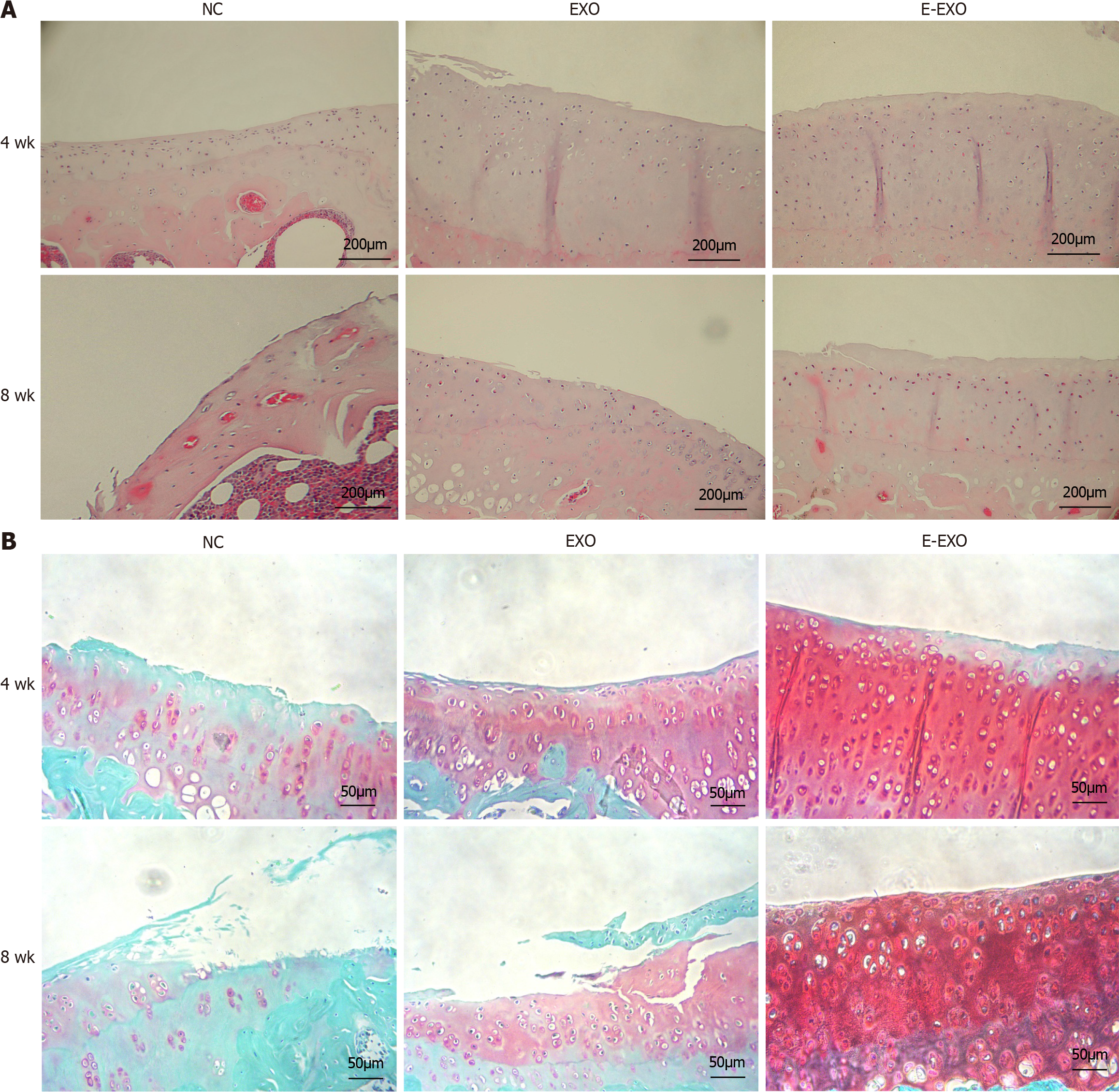
Figure 7 Images of the condyles of femur of rats stained with hematoxylin and eosin and Safranin O-fast green.
A: Images of the condyles of femur of rats stained with hematoxylin and eosin. We have re-uploaded the images; all of them depict cross-sectional staining of the femoral condyle; B: Images of condyles of femur of rats stained with Safranin O-fast green. Exo: Exosomes; NC: Negative control.
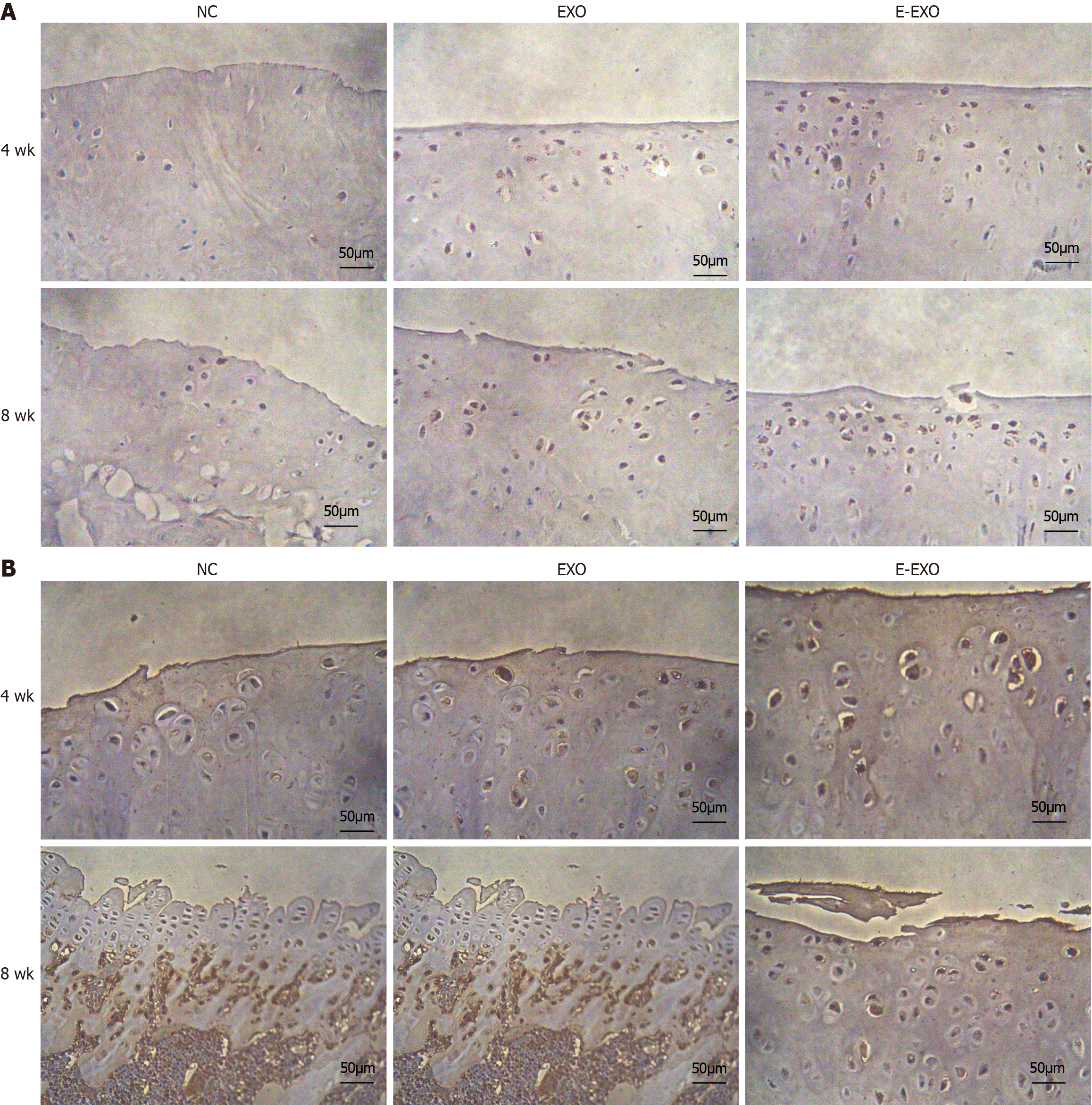
Figure 8 The expression of type II collagen and proteoglycans.
A: The expression of type II collagen; B: The expression of proteoglycans. Exo: Exosomes; NC: Negative control.
- Citation: Yang F, Xiong WQ, Li CZ, Wu MJ, Zhang XZ, Ran CX, Li ZH, Cui Y, Liu BY, Zhao DW. Extracellular vesicles derived from mesenchymal stem cells mediate extracellular matrix remodeling in osteoarthritis through the transport of microRNA-29a. World J Stem Cells 2024; 16(2): 191-206
- URL: https://www.wjgnet.com/1948-0210/full/v16/i2/191.htm
- DOI: https://dx.doi.org/10.4252/wjsc.v16.i2.191


