Copyright
©The Author(s) 2024.
World J Stem Cells. Feb 26, 2024; 16(2): 176-190
Published online Feb 26, 2024. doi: 10.4252/wjsc.v16.i2.176
Published online Feb 26, 2024. doi: 10.4252/wjsc.v16.i2.176
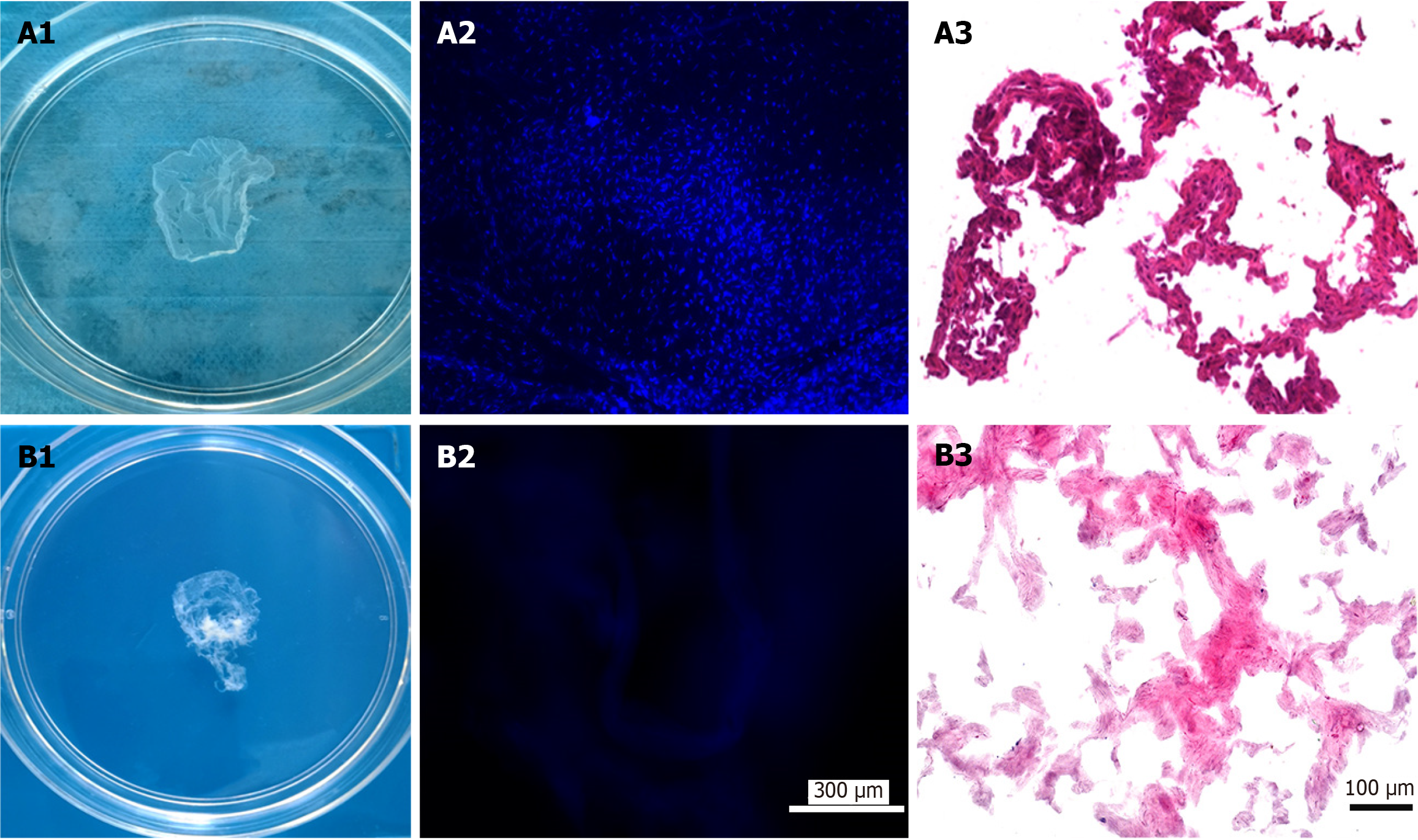
Figure 1 Fabrication of antler reserve mesenchymal cell-extracellular matrix-sheet.
A: Before decellularization: A1: Appearance of the sheet; A2: 4’,6-diamidino-2-phenylindole (DAPI) staining of the sheet; A3: Hematoxylin and eosin (HE) staining of the sheet; B: After decellularization: B1: Appearance of the sheet; B2: DAPI staining of the sheet; B3: HE staining of the sheet. Note that the decellularization process was successful and almost completely removed cells, evidenced by both DAPI and HE staining.
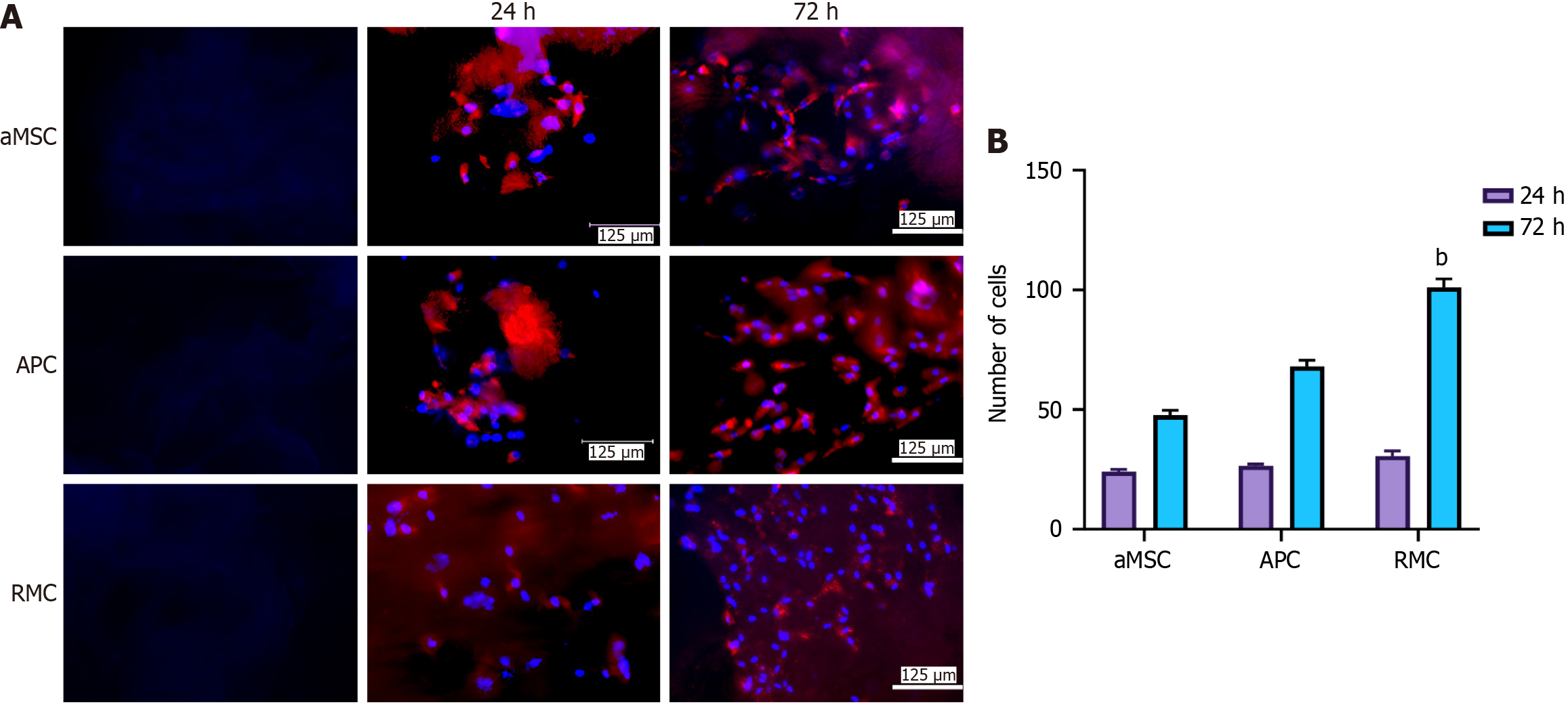
Figure 2 Effects of different extracellular matrix-sheets on cell attachment and proliferation in vitro.
A: Attachment and propagation of bone marrow mesenchymal stromal cells (bMSCs) on the fabricated adipocyte-derived MSC (aMSC-), antlerogenic periosteal cell (APC-), and antler reserve mesenchymal cell-extracellular matrix (RMC-ECM) sheets at two time points (24 and 72 h). 4’,6-diamidino-2-phenylindole (DAPI) staining: Blue and bMSCs red (pre-labelled with PKH26). First column: Decellularized ECM-sheets; second column: Cultured rat bMSCs 24 h after seeding on the sheets; third column: Cultured rat bMSCs 72 h after seeding on the sheets. Note that all three sheets were suitable for mesenchymal stromal cells to attach and proliferate; RMC-ECM-sheets not only had more cells but also less evidence of PKH26 label (red), indicating PKH26 color was more diluted due to subjecting more cycles of division; B: Quantification of cell numbers cultured on each ECM-sheet. Note that bMSCs were successfully attached and actively proliferated at 24 h after seeding, although no significant difference in cell numbers was detected amongst the three types of ECM-sheets. At 72 h, cell numbers of all groups were significantly increased compared to the corresponding group at 24 h (P < 0.001); and highly significantly differences in cell numbers were detected amongst these three groups: RMC group was higher (P < 0.01) than those of aMSC group and APC group; and APC group was significantly higher than that of aMSC group (P < 0.05). bMSCs: bone marrow mesenchymal stromal cell; aMSC: Adipocyte-derived mesenchymal stromal cell; APC: Antlerogenic periosteal cell; RMC: Antler reserve mesenchymal cell.
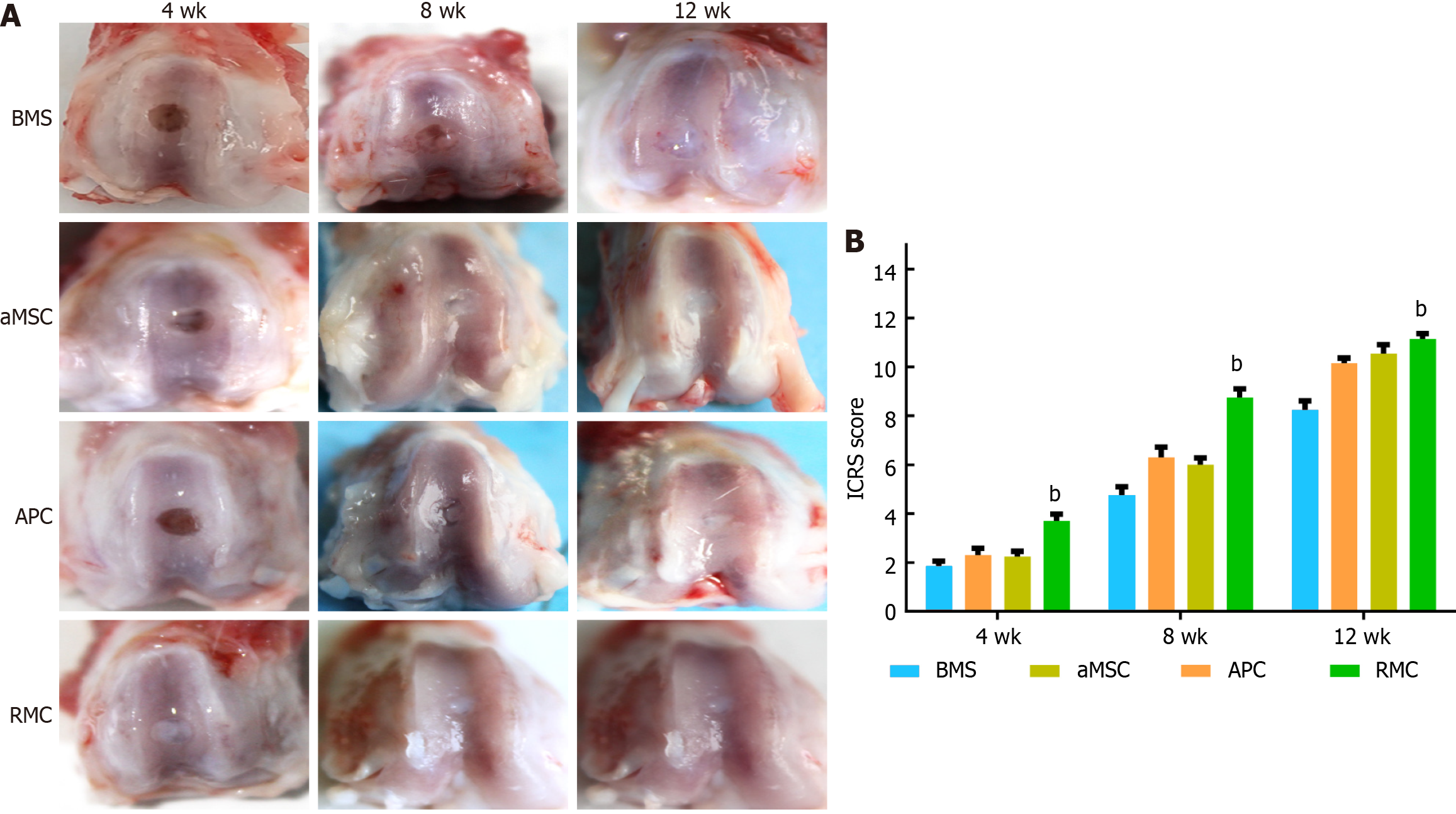
Figure 3 Morphological evaluation and International Cartilage Repair Society scores of the osteochondral defect repair by applying different adipocyte-derived mesenchymal stromal cell-, antlerogenic periosteal cell-, and antler reserve mesenchymal cell-extracellular matrix sheets at 4, 8 and 12 wk.
A: Morphological evaluation; B: International Cartilage Repair Society scores. Note that at week 4, the defect in the antler reserve mesenchymal cell (RMC) group was fully filled with ivory white colored tissue; and in the antlerogenic periosteal cell (APC) and adipocyte-derived mesenchymal stromal cell (aMSC) groups only partially filled with reddish colored tissue; in the bone marrow stimulation (BMS) group, the defect seemed empty and no regenerated tissue was visible. At week 8, the sizes of all the defects in the four groups were substantially reduced, and in the aMSC, RMC and APC groups, they were filled with white colored tissue, although the reduced defect in the BMS group was still partially filled with fibrous-like tissue. At week 12, the defect sizes of all groups were further reduced but to varying degrees, with the RMC group the smallest and BMS and aMSC groups the largest. The International Cartilage Repair Society sores were consistent with the results of morphological observation. bP < 0.01. BMS: Bone marrow stimulation; aMSC: Adipocyte-derived mesenchymal stromal cell; APC: Antlerogenic periosteal cell; RMC: Antler reserve mesenchymal cell.
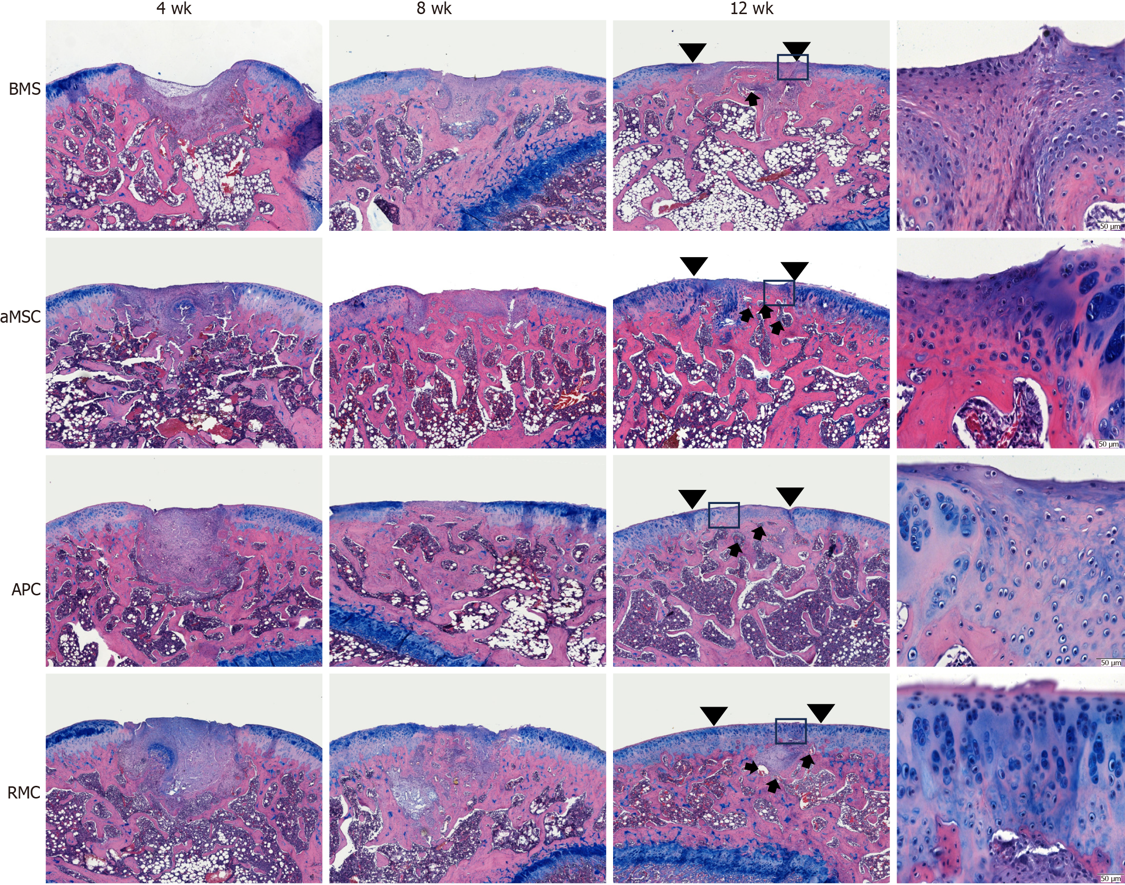
Figure 4 Histological evaluation using counter-staining of hematoxylin and eosin and alcian blue for osteochondral defect repair by applying different extracellular matrix-sheets at 4, 8 and 12 wk.
Insets are the enlarged area for more detailed evaluation of critical sites at week 12. Note that, consistent with the morphological evaluation, histologically at week 4, the defects in the bone marrow stimulation (BMS) group had only started to be filled with fibrous tissue, whereas those of the other three groups were fully filled with the regenerated tissue. The filled tissue in antler reserve mesenchymal cell (RMC) group was stained bluer (proteoglycan) by alcian blue, indicating that the tissue was more cartilage in nature. At week 8, the repair process had almost reached completion except for the BMS group, although the regenerated tissue varied in type among the different groups. In the BMS group, fibrous tissue with negligible osseous tissue was formed; in the adipocyte-derived mesenchymal stromal cell (aMSC) group, osseous tissue with a rough surface and with negligible fibrous tissue was formed; in the antlerogenic periosteal cell (APC) group, only osseous tissue with a smooth surface had formed; in the RMC group, osteocartilaginous tissue was detected with cartilage tissue evident on the outermost surface. At week 12, the repair process was essentially complete, although the quality of the repair varied greatly among the groups. In the BMS group, a mixture of osseous and fibrous tissue in the defect (arrowheads) was formed with the fibrous tissue replacing the cartilage as a surface layer (inset); in both the aMSC and APC groups, the reconstructed cartilage layer (arrowheads; fibrous in nature) was not as thick as the original, and resident chondrocytes were randomly distributed (inset), although the APC group had a smoother surface than aMSC group; in the RMC group, the osteochondral defect was perfectly repaired with a well-reconstructed smooth-surfaced outermost layer of cartilage (arrowheads; hyaline in nature), and the resident chondrocytes were arranged in a stacked style, typical of articular cartilage, see inset). Bar = 500 μm. BMS: Bone marrow stimulation; aMSC: Adipocyte-derived mesenchymal stromal cell; APC: Antlerogenic periosteal cell; RMC: Antler reserve mesenchymal cell.
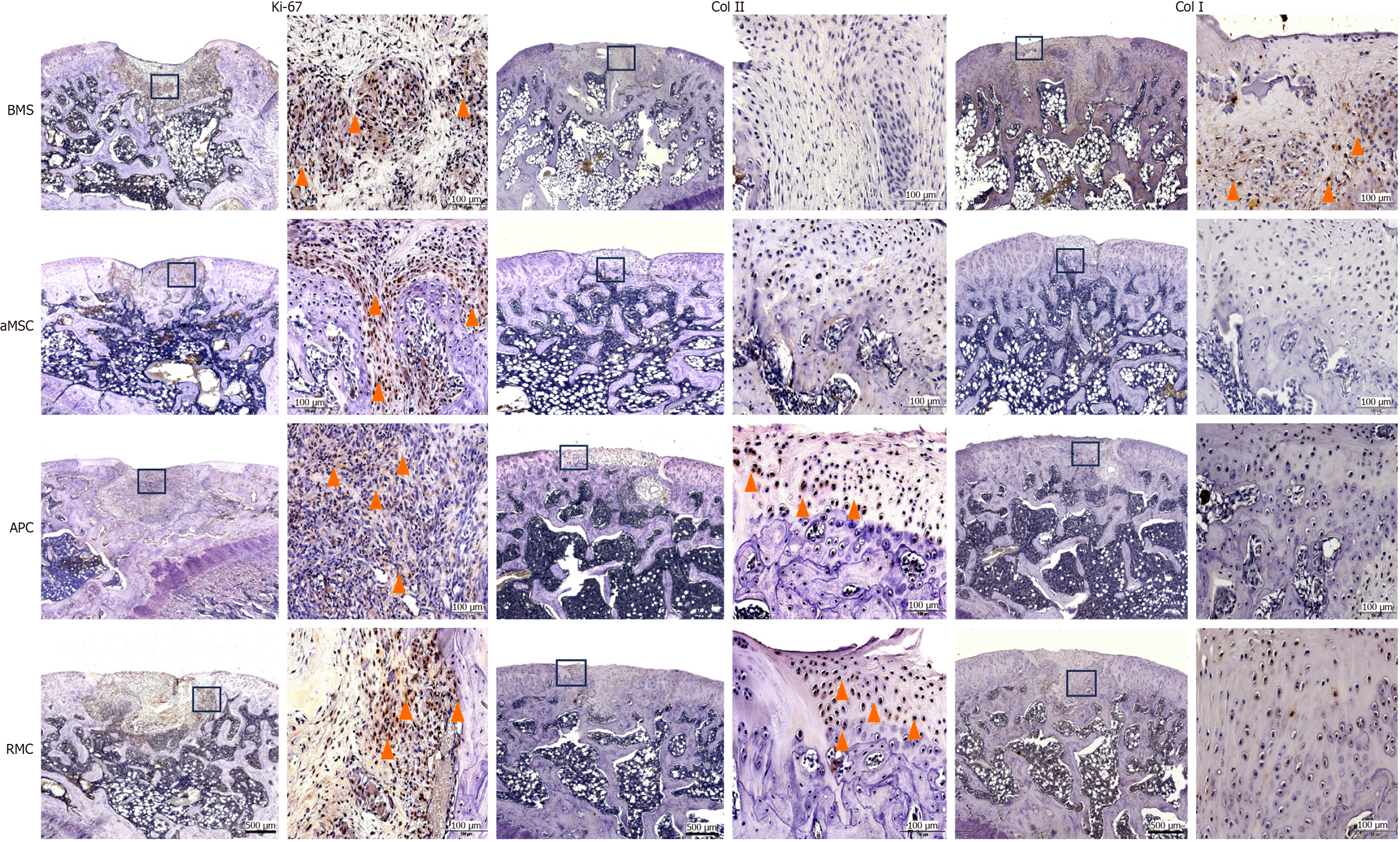
Figure 5 Immunohistochemical evaluation of the top layer covering the osteochondral defects by applying different extracellular matrix-sheets.
Ki-67 staining at week 4: Note that the cells of the re-forming tissue in the defects of all groups were extensively and intensively stained with Ki-67 (yellow arrowheads), indicating that each defect was under active repair. Col II staining at week 12: Note that the surface layer of each defect was positively stained in the antlerogenic periosteal cell (APC) and antler reserve mesenchymal cell (RMC) groups only (yellow arrowheads), but not in the adipocyte-derived mesenchymal stromal cell (aMSC) and bone marrow stimulation (BMS) groups, indicating that the surface tissue in the APC and RMC groups was of cartilage in nature. Col I staining at week 12: Note that the regenerated tissue in the BMS group was significantly positively stained (yellow arrowheads), but that in the other three groups was not, indicating that the surface tissue in the BMS group was more fibrous tissue-like in nature. Bar = 500 μm. BMS: Bone marrow stimulation; aMSC: Adipocyte-derived mesenchymal stromal cell; APC: Antlerogenic periosteal cell; RMC: Antler reserve mesenchymal cell.
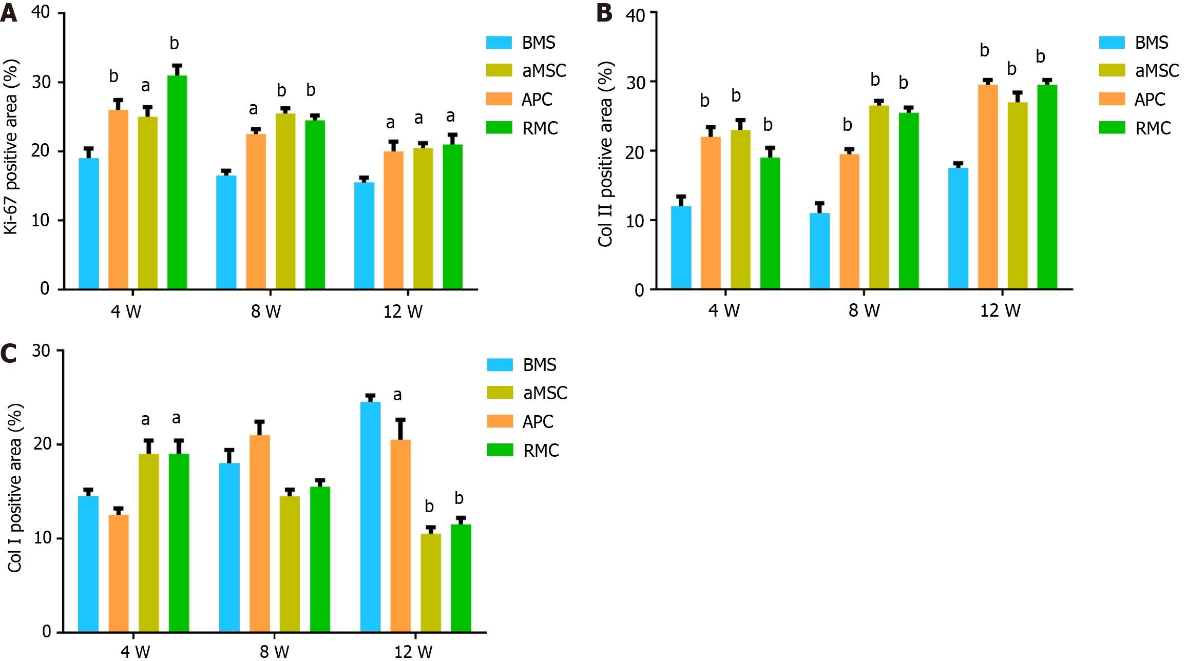
Figure 6 Quantification (%) of immunohistochemical positive staining.
A-C: The overall results were consistent with those of Figure 5. Ki-67 staining: The reserve mesenchymal cell (RMC) group was the highest at week 4, but gradually decreased over time; at weeks 8 and 12, three treatment groups reached lowest level, although all were still significantly higher than the control bone marrow stimulation (BMS) group, indicating that at week 12, the healing was almost complete. Col II staining: The BMS group was the lowest at 4, 8 and 12 wk, with the three treatment groups reaching a similar level at week 12, indicating that the treatment had greatly boosted cartilage formation. Col I staining: At week 4, the antlerogenic periosteal cell (APC) and RMC groups were significantly higher than the adipocyte-derived mesenchymal stromal cell (aMSC) and the control BMS groups, but at weeks 8 and 12 were significantly lower than that of the aMSC and the BMS groups; indicating that the two deer groups contained less bone or fibrous tissue. aP < 0.05; bP < 0.01. BMS: Bone marrow stimulation; aMSC: Adipocyte-derived mesenchymal stromal cell; APC: Antlerogenic periosteal cell; RMC: Antler reserve mesenchymal cell.
- Citation: Wang YS, Chu WH, Zhai JJ, Wang WY, He ZM, Zhao QM, Li CY. High quality repair of osteochondral defects in rats using the extracellular matrix of antler stem cells. World J Stem Cells 2024; 16(2): 176-190
- URL: https://www.wjgnet.com/1948-0210/full/v16/i2/176.htm
- DOI: https://dx.doi.org/10.4252/wjsc.v16.i2.176


