Copyright
©The Author(s) 2023.
World J Stem Cells. May 26, 2023; 15(5): 421-437
Published online May 26, 2023. doi: 10.4252/wjsc.v15.i5.421
Published online May 26, 2023. doi: 10.4252/wjsc.v15.i5.421
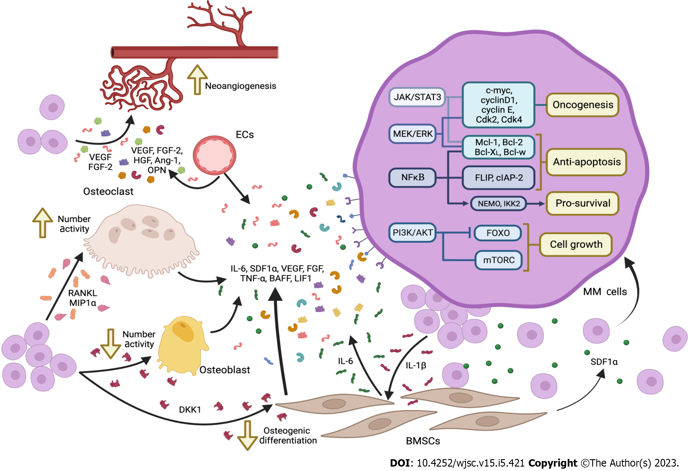
Figure 1 Schematic representation of the main factors involved in the bidirectional communication between multiple myeloma cells and cells in the bone marrow microenviroment (bone marrow mesenchymal stem cells, osteoclasts, osteoblast, etc.
). The main signaling patwthays activated by these factors are also depicted (Created with Biorender.com). VEGF: Vascular endothelial growth factor; FGF: Fibroblast growth factors; HGF: Hepatocyte growth factor; OPN: Osteopontin; ECs: Endothelial cells; IL: Interleukin; SDF1α: Stromal cell derived factor 1α; TNF-α: Tumor necrosis factor-α; BAFF: B-cell activating factor; DKK-1: Dickkopf-1; MM: Multiple myeloma; BM-MSC: Bone marrow mesenchymal stem cells; JAK: Janus kinase; STAT3: Signal transducer and activator of transcription 3; NFκΒ: Nuclear factor kappa-Β; PI3K: Phosphatidylinositol 3-kinase; RANKL: Receptor activator of NFκΒ ligand; Ang-1: angiopoietin-1; MEK: MAPK kinase; ERK: Extracellular signal regulated kinase; LIF1: Leukemia inhibitory factor-1. Created with BioRender.com.
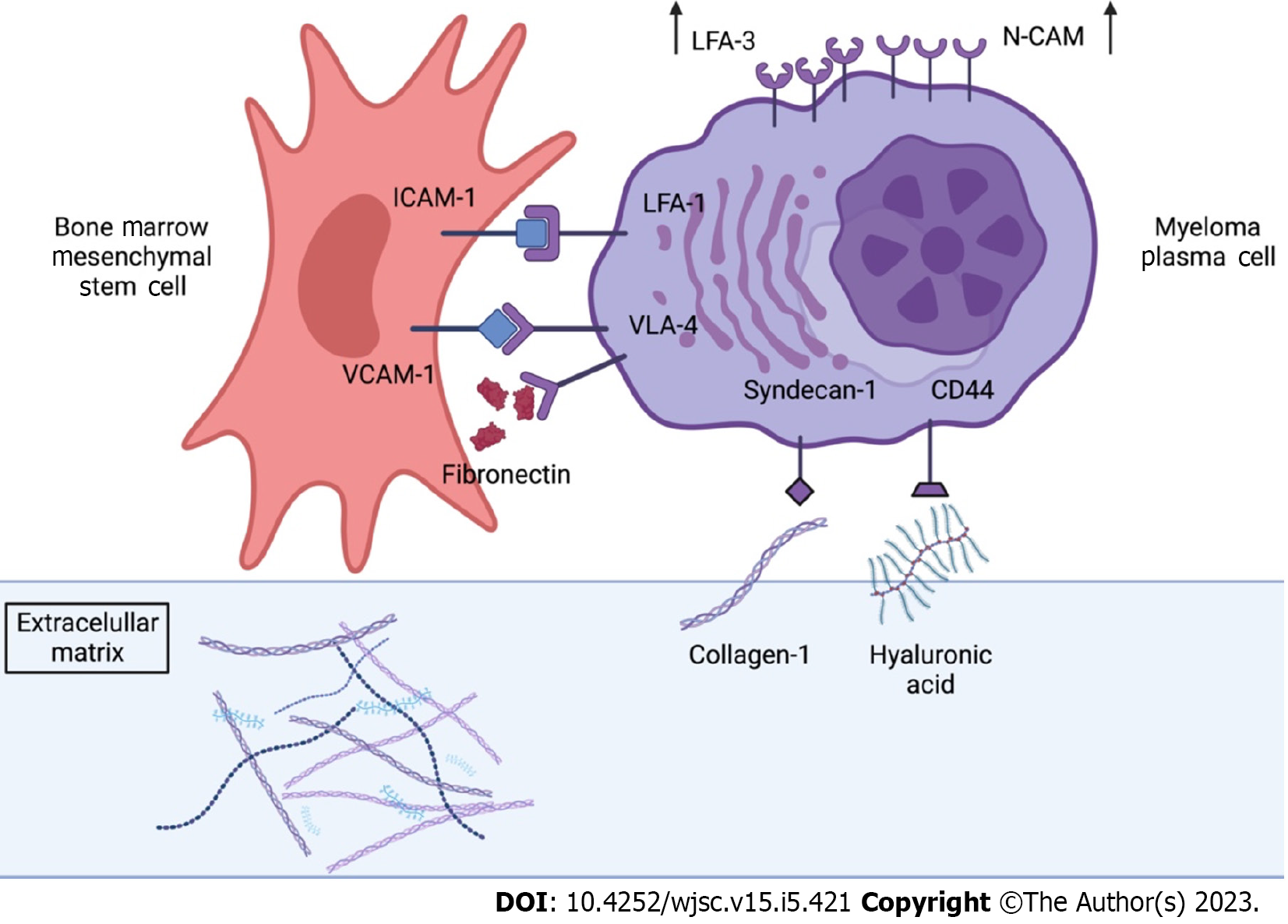
Figure 2 Schematic representation of the main cell adhesion molecules in multiple myeloma cells and bone marrow mesenchymal stem cells.
The main interactions between cell adhesion molecules (CAMs) of these two types of cells as well as the interactions of these CAMs with proteins of the extracellular matrix are displayed (Created with Biorender.com). ICAM-1: Intercellular adhesion molecule 1; VCAM-1: Vascular cell adhesion molecule-1; LFA-1: Leukocyte function-associated antigen 1; N-CAM: Neural cell adhesion molecule; VLA: Very late antigen. Created with BioRender.com.
- Citation: García-Sánchez D, González-González A, Alfonso-Fernández A, Del Dujo-Gutiérrez M, Pérez-Campo FM. Communication between bone marrow mesenchymal stem cells and multiple myeloma cells: Impact on disease progression. World J Stem Cells 2023; 15(5): 421-437
- URL: https://www.wjgnet.com/1948-0210/full/v15/i5/421.htm
- DOI: https://dx.doi.org/10.4252/wjsc.v15.i5.421


