Copyright
©The Author(s) 2023.
World J Stem Cells. Dec 26, 2023; 15(12): 1077-1092
Published online Dec 26, 2023. doi: 10.4252/wjsc.v15.i12.1077
Published online Dec 26, 2023. doi: 10.4252/wjsc.v15.i12.1077
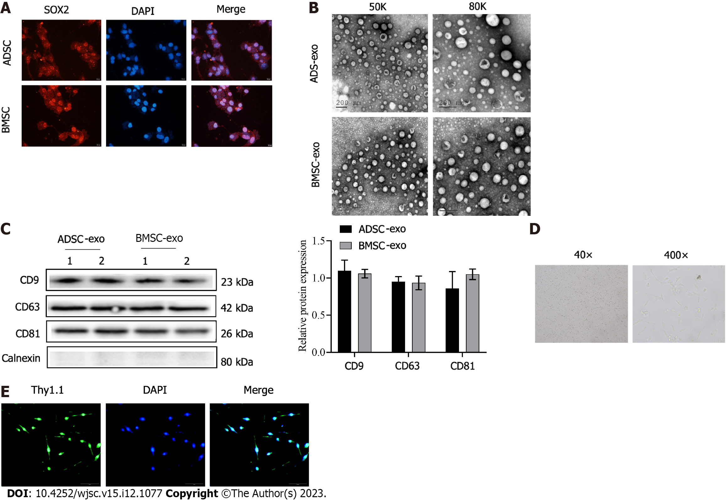
Figure 1 Adipose-derived stem cell and bone marrow-derived stem cell-exosomes, and retinal ganglion cells were isolated and identified.
A: The immunofluorescence staining of stem cell markers SOX2 (Scale bar: 20 μm); B: The morphology of adipose-derived stem cell-exosomes (ADSC-Exos) and bone marrow-derived stem cell (BMSC)-Exos was observed by electron microscopy (scale bar: 200 nm); C: The ADSC-Exos and BMSC-Exos marker molecules (CD9, CD63, and CD81) were detected by western blotting; D: The light microscope images of retinal ganglion cells (RGCs) (magnification: 40 × and 400 ×); E: The isolated RGCs was examined by Thy1.1 immunofluorescence (scale bar: 50 μm). ADSC: Adipose-derived stem cell; BMSC: Bone marrow-derived stem cell; Exo: Exosome.
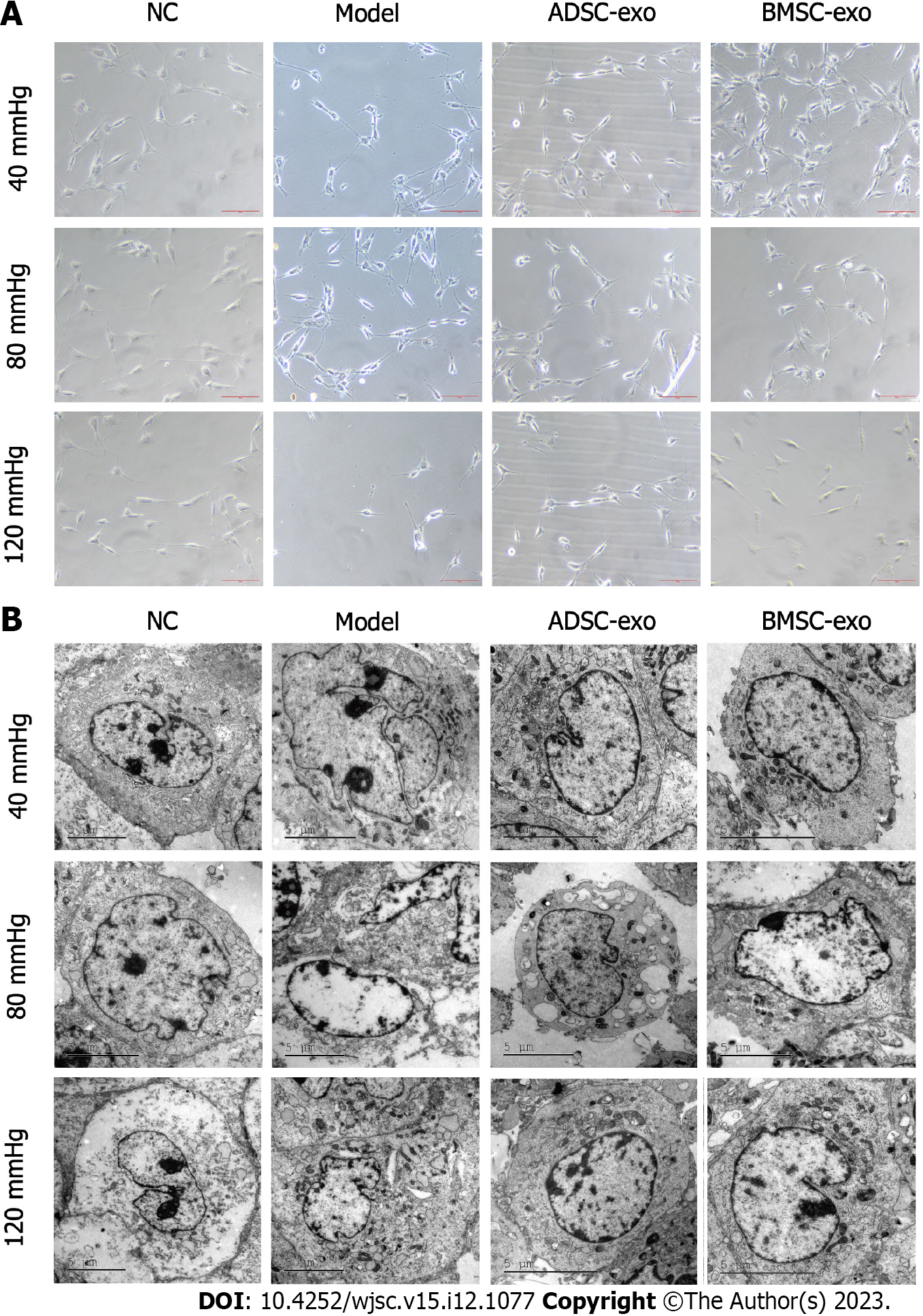
Figure 2 The growth of retinal ganglion cells.
A: The observation of morphology and growth of retinal ganglion cells (RGCs) by phase contrast microscope (Scale bar: 50 μm); B: The observation of RGCs morphology by transmission electron microscope (Scale bar: 5 μm). ADSC: Adipose-derived stem cell; BMSC: Bone marrow-derived stem cell; Exo: Exosome; NC: Normal control.
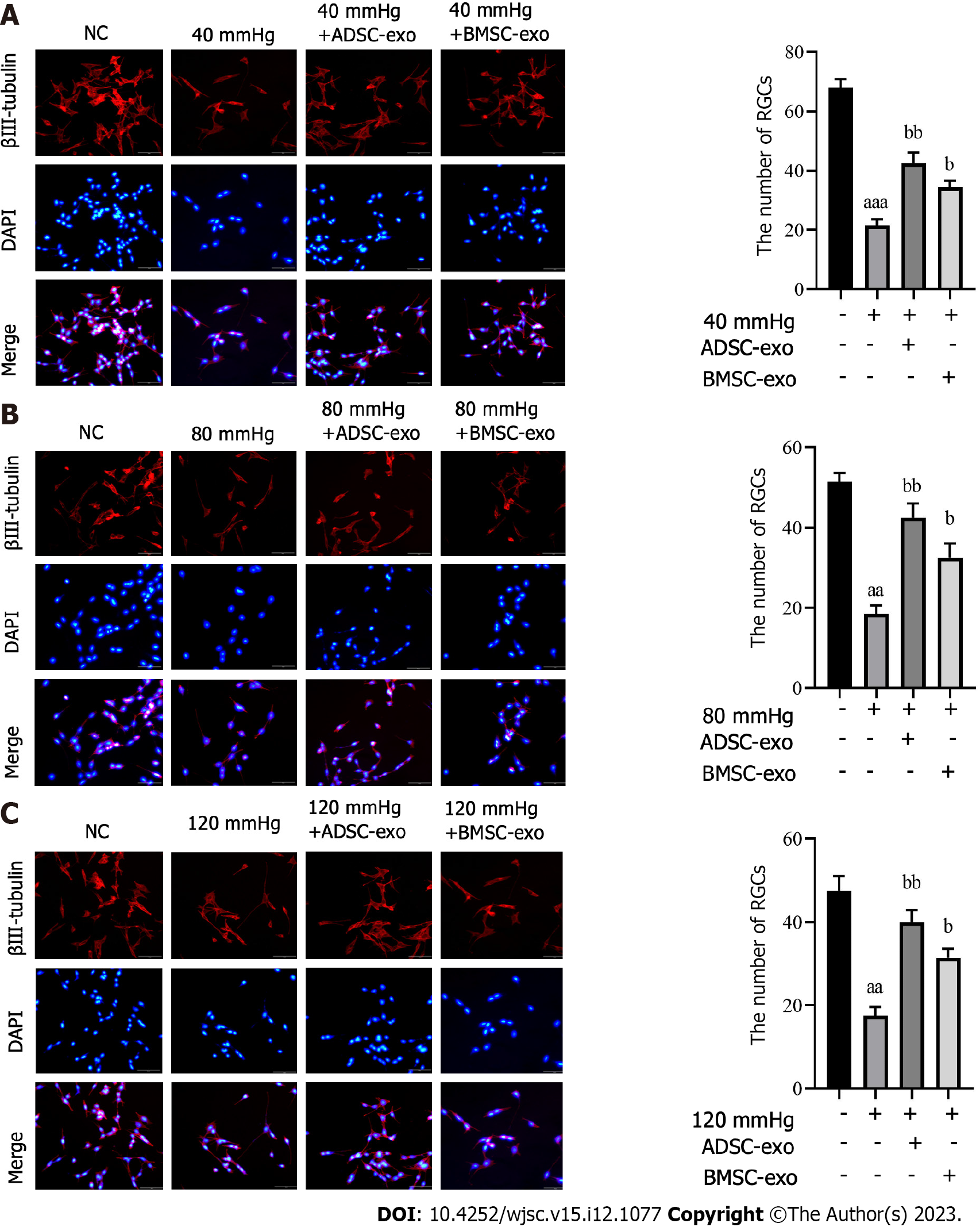
Figure 3 Immunofluorescence detection of β-III tubulin.
A-C: Immunofluorescence staining of β-III tubulin (red) and nuclear staining (DAPI, blue) (left panel) and the number of β-III tubulin-positive retinal ganglion cells (right panel) in different groups after exposure to 40 mmHg (A), 80 mmHg (B), and 120 mmHg (C). aP < 0.05, compared with the normal control group (scale bar: 50 μm); bP < 0.05, compared with model (40, 80, and 120 mmHg) group. ADSC: Adipose-derived stem cell; BMSC: Bone marrow-derived stem cell; Exo: Exosome; NC: Normal control; CREB: Cyclic adenosine monophosphate response element-binding protein.
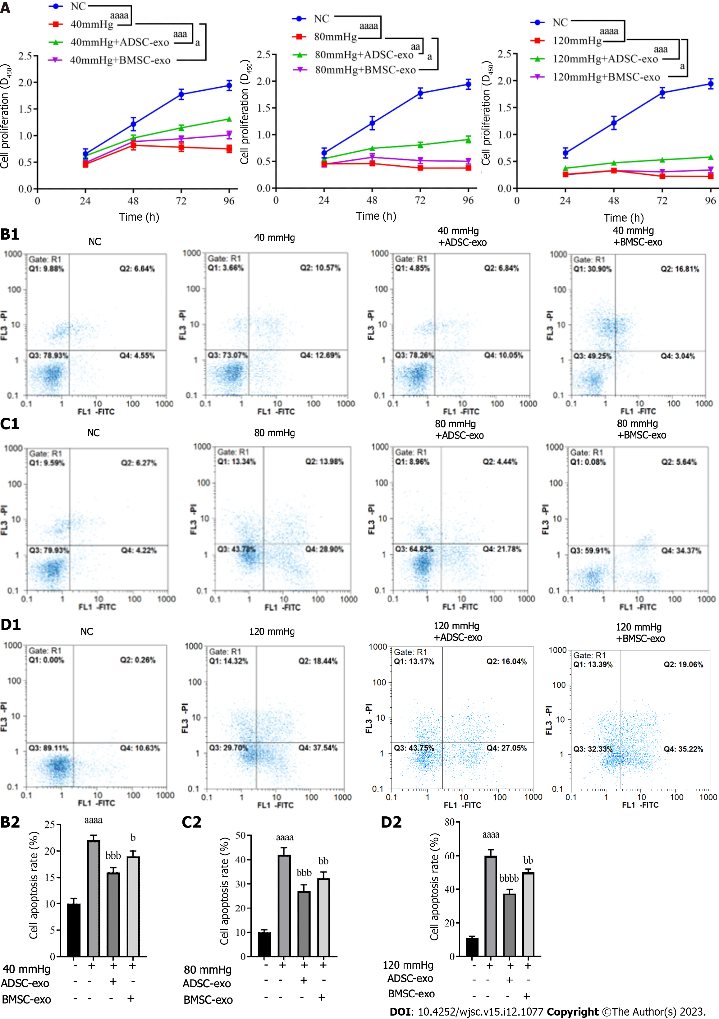
Figure 4 Retinal ganglion cells proliferation and apoptosis.
A: Cell proliferation was detected by using the Cell Counting Kit-8 proliferation reagent; B-D: Cell apoptosis was detected by flow cytometry for apoptosis detection, and representative flow cytometry density plots (left) and statistical bar chart (right). aP < 0.05, compared with the normal control group; bP < 0.05, compared with model (40, 80, and 120 mmHg) group. ADSC: Adipose-derived stem cell; BMSC: Bone marrow-derived stem cell; Exo: Exosome; NC: Normal control.
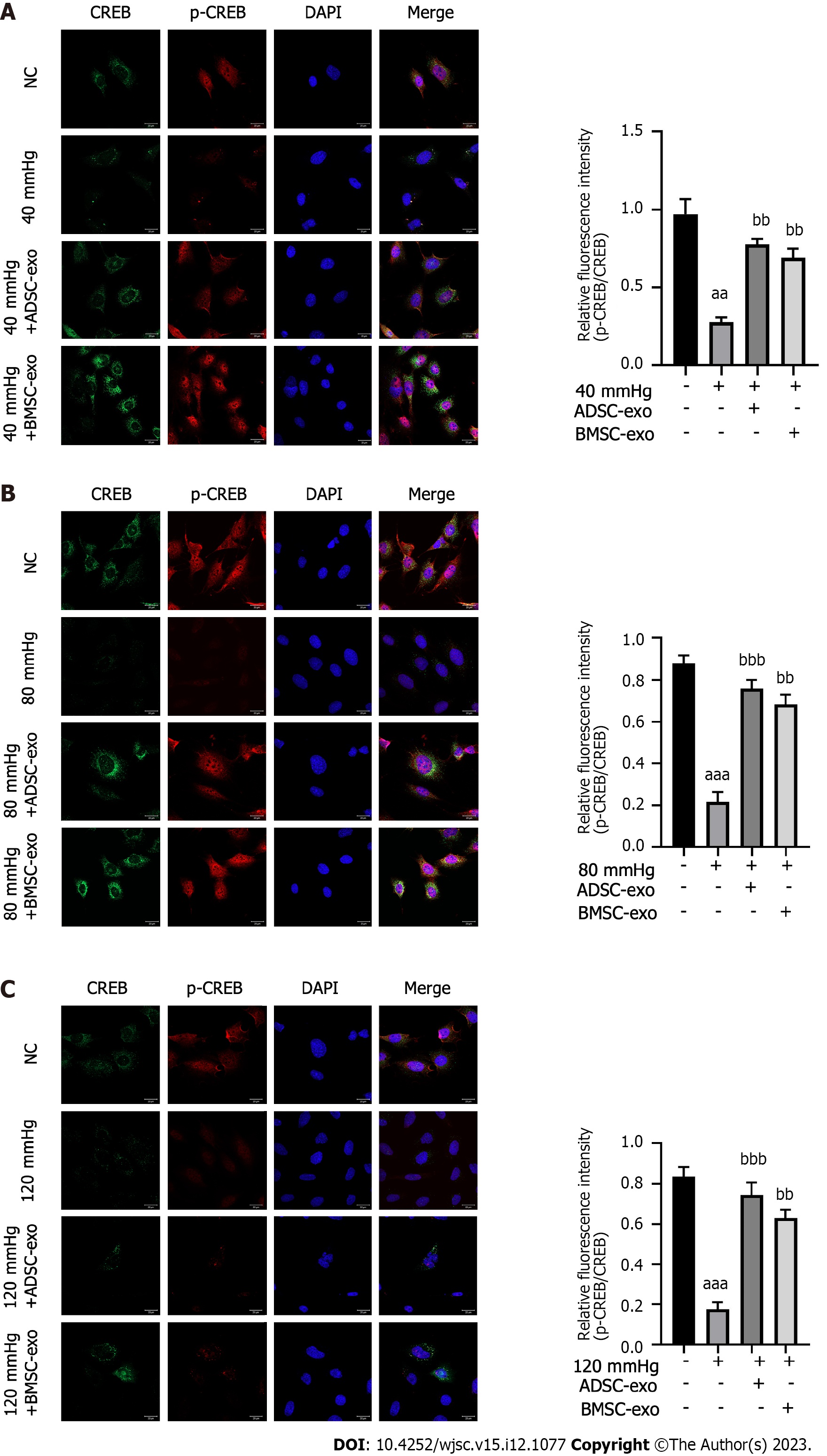
Figure 5 Immunofluorescence detection of p-CREB and CREB.
A-C: Immunofluorescence staining of CREB (green), p-CREB (red) and nuclear staining (DAPI, blue) (left panel) and quantification of the ratio of p-CREB/CREB (right panel) in different groups after exposure to (A) 40 mmHg, (B) 80 mmHg, and (C) 120 mmHg (Scale bar: 20 μm). aP < 0.05, compared with the normal control group; bP < 0.05, compared with the model (40, 80, and 120 mmHg) group. ADSC: Adipose-derived stem cell; BMSC: Bone marrow-derived stem cell; Exo: Exosome; NC: Normal control.
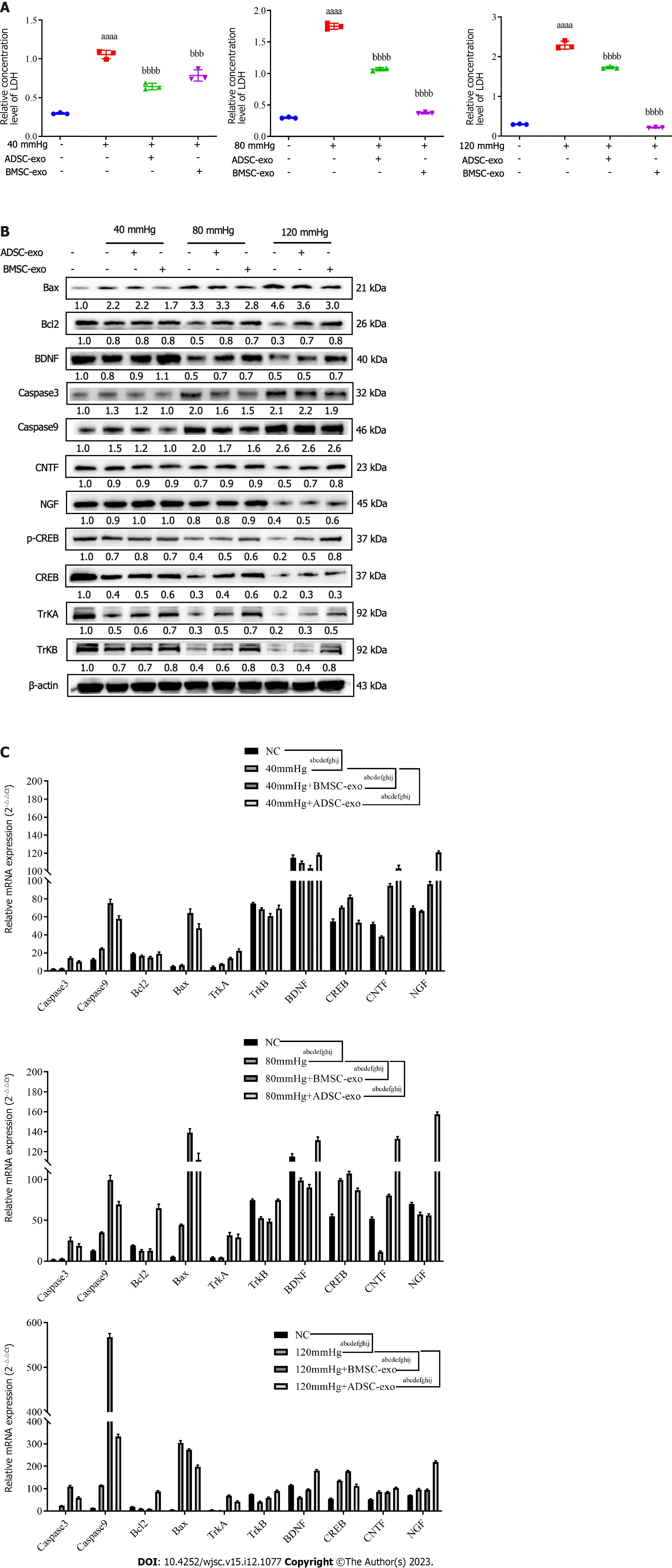
Figure 6 The expression of lactate dehydrogenase, neurotrophic factor and apoptosis-related protein and mRNA.
A: Lactate dehydrogenase (LDH) expression level in each group was measured by the LDH test kit; B: The expression of apoptosis-related proteins and neurotrophic factors were detected by western blotting; C: The mRNA expression of apoptosis-related proteins and neurotrophic factors were detected by real-time quantitative polymerase chain reaction. aP, bP, cP, dP, eP, fP, gP, hP, iP, and jP < 0.05, represent the comparison of caspase3, caspase9, B-cell lymphoma 2, Bax, tropomyosin receptor kinase (TrK)A, TrKB, brain-derived neurotrophic factor, cyclic adenosine monophosphate response element-binding protein, ciliary neurotrophic factor, and nerve growth factor proteins between the two groups. ADSC: Adipose-derived stem cell; BMSC: Bone marrow-derived stem cell; Exo: Exosome; NC: Normal control; LDH: Lactate dehydrogenase; Bcl2: polyvinylidene fluoride; Trk: Tropomyosin receptor kinase; BDNF: Brain-derived neurotrophic factor; CREB: Cyclic adenosine monophosphate response element-binding protein; CNTF: Ciliary neurotrophic factor; NGF: Nerve growth factor.
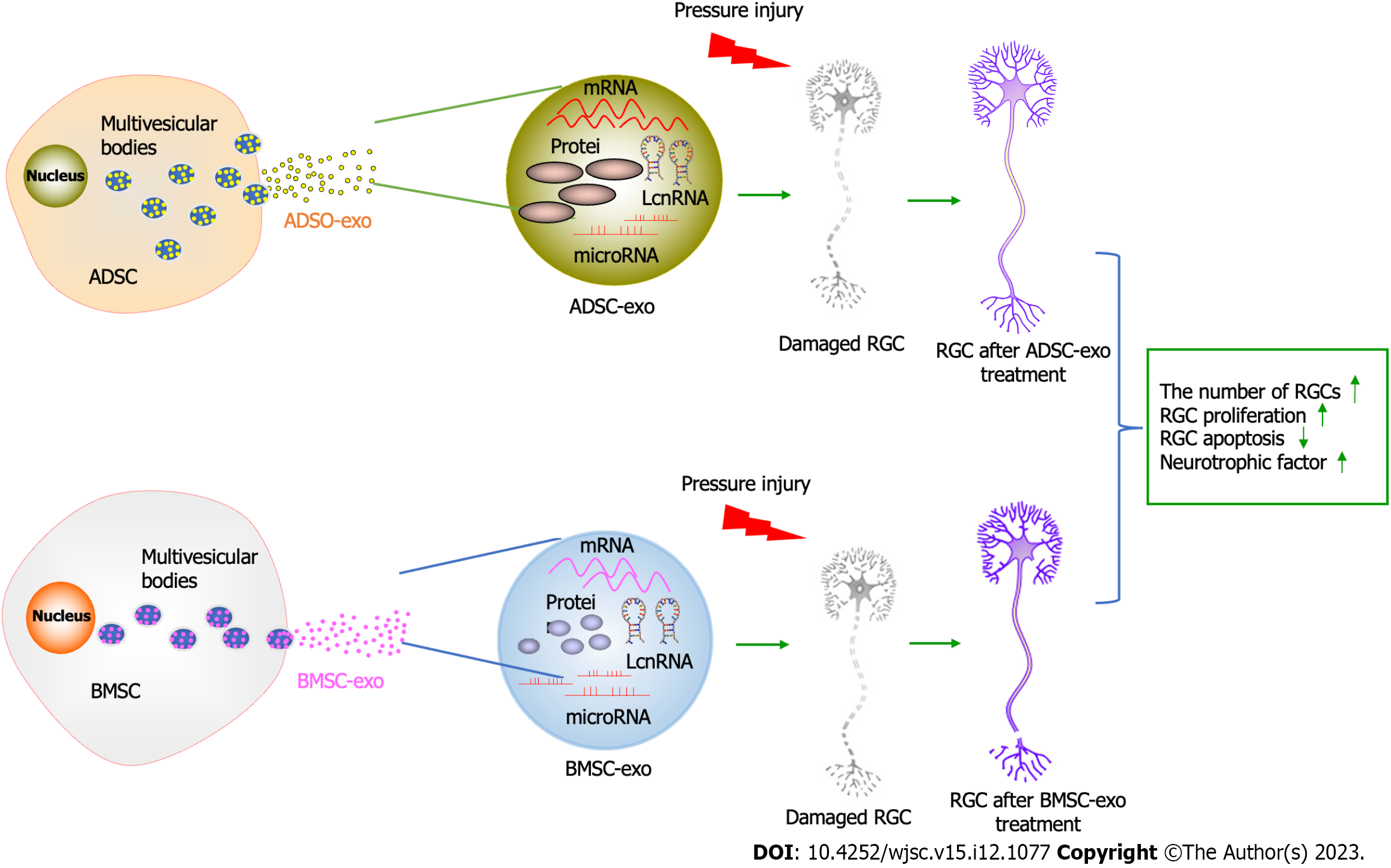
Figure 7 Graphical abstract.
ADSC: Adipose-derived stem cell; BMSC: Bone marrow-derived stem cell; LncRNA: Long non-coding RNA; RGC: Retinal ganglion cell; Exo: Eexosome.
- Citation: Zheng ZK, Kong L, Dai M, Chen YD, Chen YH. ADSC-Exos outperform BMSC-Exos in alleviating hydrostatic pressure-induced injury to retinal ganglion cells by upregulating nerve growth factors. World J Stem Cells 2023; 15(12): 1077-1092
- URL: https://www.wjgnet.com/1948-0210/full/v15/i12/1077.htm
- DOI: https://dx.doi.org/10.4252/wjsc.v15.i12.1077


