Copyright
©The Author(s) 2020.
World J Stem Cells. Jun 26, 2020; 12(6): 514-526
Published online Jun 26, 2020. doi: 10.4252/wjsc.v12.i6.514
Published online Jun 26, 2020. doi: 10.4252/wjsc.v12.i6.514
Figure 1 Flow chart of selection criteria.
MCOA: Medial compartment osteoarthritis; MFC: Medial femoral condyle; HTO: High tibial osteotomy; hUCB-MSCs: Human umbilical cord blood-derived mesenchymal stem cells.
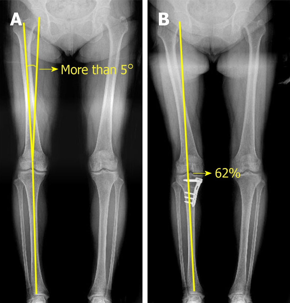
Figure 2 High tibial osteotomy.
A: High tibial osteotomy was performed at a hip-knee-ankle angle of 5° or more; B: The mechanical axis was corrected to approximately 62% lateral to the tibial plateau.
Figure 3 Arthroscopic findings of stem cell implantation procedures.
A: Medial compartment osteoarthritis (arrow) in a 61-year-old woman; B: Multiple holes, 4 mm in diameter and 4 mm in depth (arrow), were drilled using a drill bit; C: Human umbilical cord blood-derived mesenchymal stem cells were mixed with hyaluronic acid hydrogel and implanted in the holes (arrow).
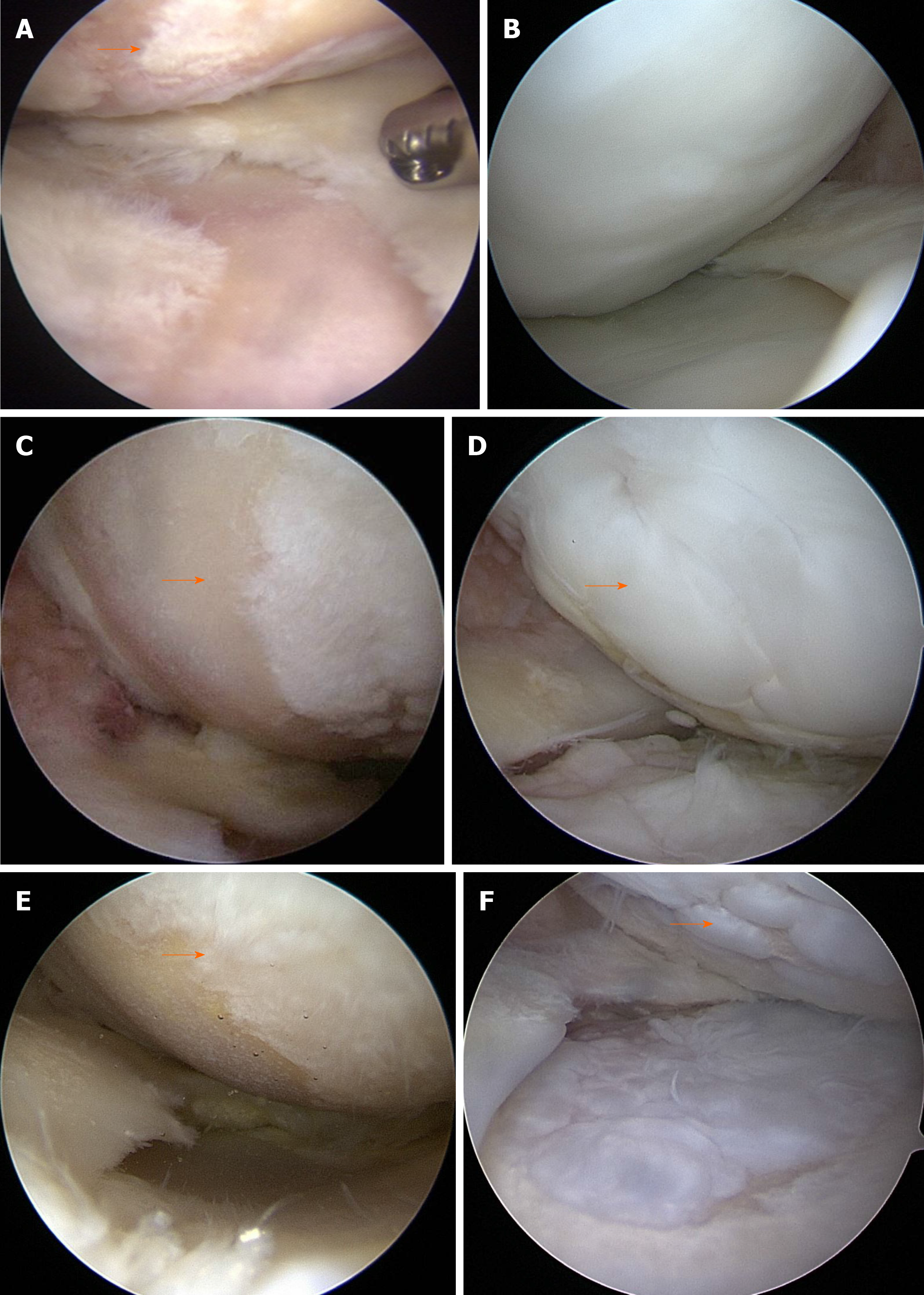
Figure 4 Second-look arthroscopic findings.
A: Cartilage lesions classified as International Cartilage Repair Society (ICRS) grade IV in the medial femoral condyle (MFC) (arrow) and tibial plateau in a 61-year-old female patient; B: Cartilage was regenerated to ICRS grade I (arrow) via second-look arthroscopy, performed 13 mo after the initial operation; C: A cartilage lesion classified as ICRS grade IV in the MFC (arrow) of a 52-year-old male patient; D: Cartilage was regenerated to ICRS grade II (arrow) via second-look arthroscopy, performed 22 mo after the initial operation; E: A cartilage lesion of ICRS grade IV in the MFC (arrow) of a 68-year-old female patient; F: Cartilage was regenerated to ICRS grade III (arrow) via second-look arthroscopy, performed 16 mo after the initial operation.
- Citation: Song JS, Hong KT, Kong CG, Kim NM, Jung JY, Park HS, Kim YJ, Chang KB, Kim SJ. High tibial osteotomy with human umbilical cord blood-derived mesenchymal stem cells implantation for knee cartilage regeneration. World J Stem Cells 2020; 12(6): 514-526
- URL: https://www.wjgnet.com/1948-0210/full/v12/i6/514.htm
- DOI: https://dx.doi.org/10.4252/wjsc.v12.i6.514


