Copyright
©The Author(s) 2020.
World J Stem Cells. Dec 26, 2020; 12(12): 1492-1510
Published online Dec 26, 2020. doi: 10.4252/wjsc.v12.i12.1492
Published online Dec 26, 2020. doi: 10.4252/wjsc.v12.i12.1492
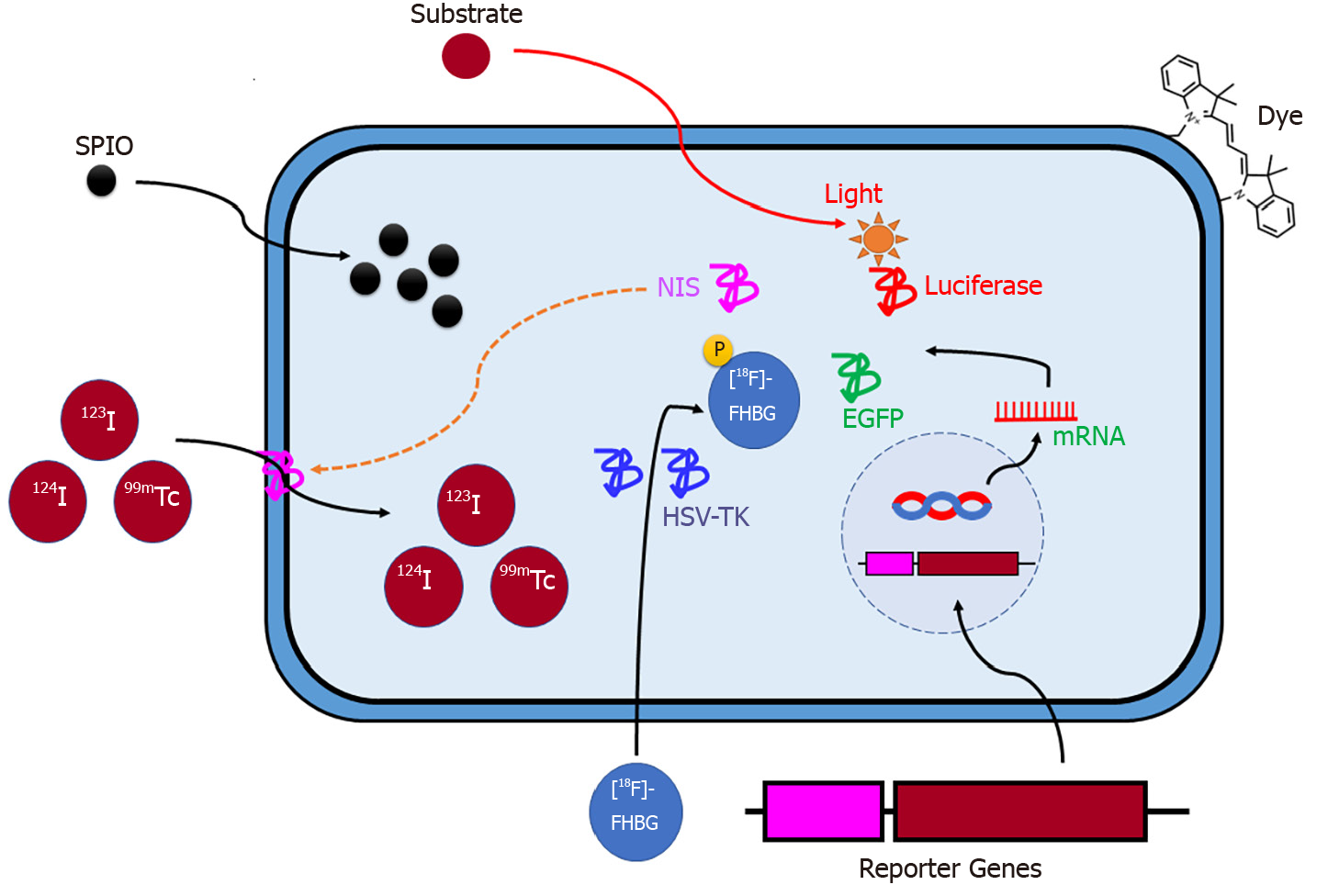
Figure 1 Schematic illustration of labeling mesenchymal stem cells for in vivo non-invasive imaging.
SPIO: Small superparamagnetic iron oxide; NIS: Sodium iodide symporter; HSV-TK: Herpes simplex virus-thymidine kinase; EGFP: Enhanced green fluorescent protein; [18F]FHBG: 9-(4-[F]fluoro-3-hydroxymethylbutyl) guanine.
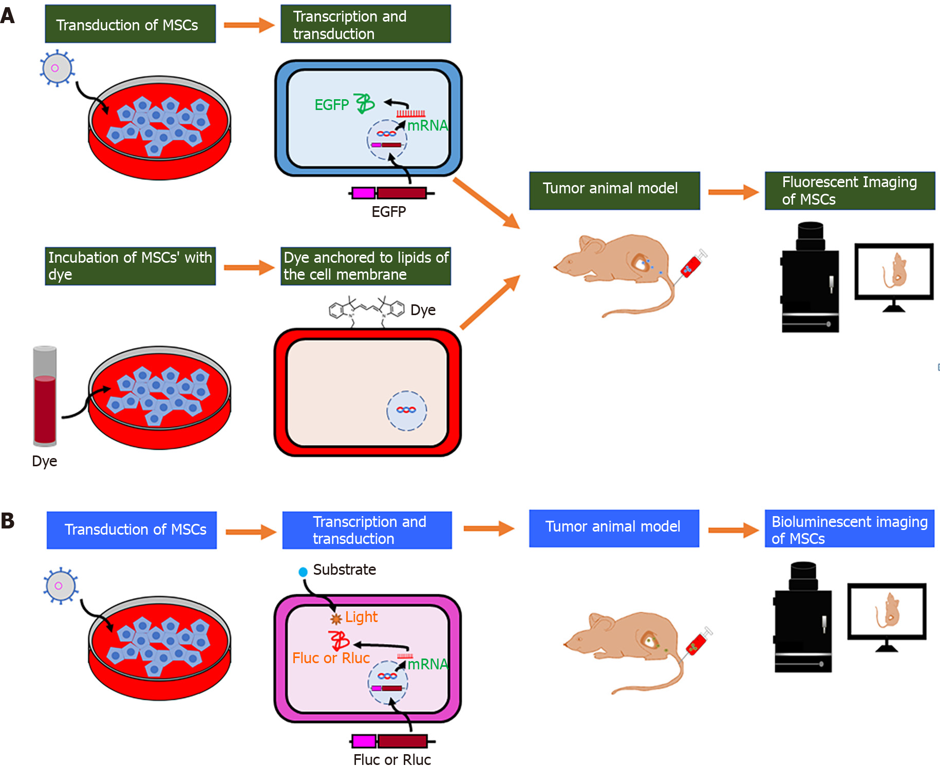
Figure 2 Schematic illustration of the labeling strategy for in vivo tracking of mesenchymal stem cells by optical imaging.
A: After fluorescent protein (enhanced green fluorescent protein) transduction into mesenchymal stem cells (MSCs) or binding of lipophilic labeling agents (e.g., fluorescent nanoparticles and VivoTrack 680) to the membrane of MSCs, cells are injected into the tumor-bearing mice, and their migration is visualized with the use of in vivo fluorescent imaging; B: After the bioluminescent protein (Firefly or Renilla luciferase) transduction into MSCs, cells are injected into the tumor-bearing mice. The light emitted due to the interaction between luciferase and its substrates (D-luciferin or coelenterazine) is captured by in vivo bioluminescent imaging. MSCs: Mesenchymal stem cells; EGFP: Enhanced green fluorescent protein; Fluc: Firefly luciferase; Rluc: Renilla luciferase.
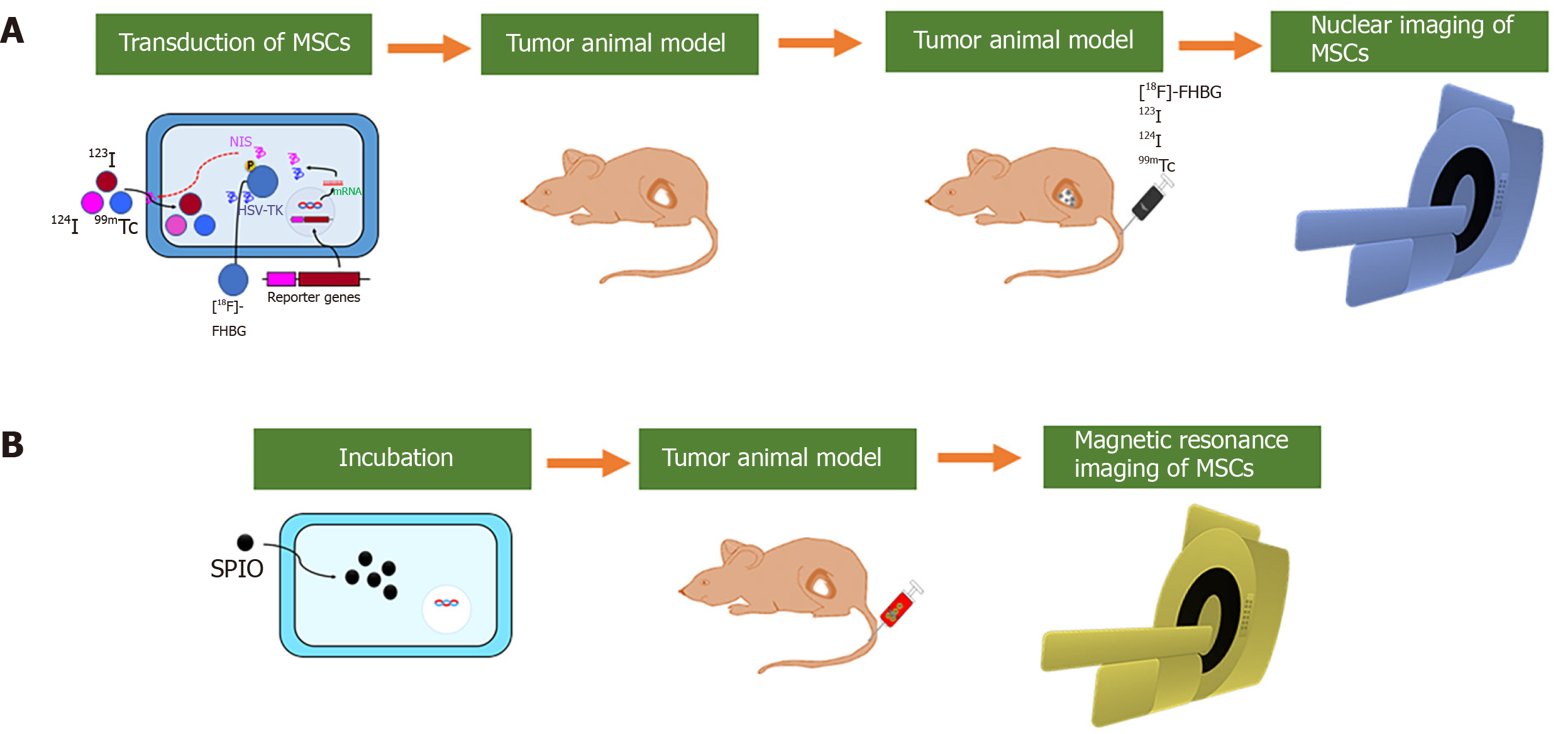
Figure 3 Schematic illustration of the labeling strategy for in vivo tracking of mesenchymal stem cells by nuclear and magnetic resonance imaging.
A: Gene transduction of sodium iodide symporter (NIS) or herpes simplex virus-thymidine kinase (HSV-TK) into mesenchymal stem cells (MSCs) can aid radiotracers (123I, 124I and 99mTc) in entering MSCs. MSCs-NIS are injected into tumor-bearing mice followed by the injection of radiotracers. In vivo nuclear imaging (positron-emission tomography, camera imaging, and single-photon emission computed tomography) can visualize migration of the MSCs; B: MSCs can be incubated with molecules including small superparamagnetic iron oxide (SPIO) or SPIO coated with gold-nanoparticles (SPIO@Au-NPs). SPIO-labeled MSCs are injected into tumor-bearing mice, and in vivo magnetic resonance imaging can visualize migration of the MSCs. MSCs: Mesenchymal stem cells; NIS: Sodium iodide symporter; HSV-TK: Herpes simplex virus-thymidine kinase; 99mTc: Technetium-99m; [18F]FHBG: 9-(4-[F]fluoro-3-hydroxymethylbutyl) guanine; SPIO: Superparamagnetic iron oxide.
- Citation: Rajendran RL, Jogalekar MP, Gangadaran P, Ahn BC. Noninvasive in vivo cell tracking using molecular imaging: A useful tool for developing mesenchymal stem cell-based cancer treatment. World J Stem Cells 2020; 12(12): 1492-1510
- URL: https://www.wjgnet.com/1948-0210/full/v12/i12/1492.htm
- DOI: https://dx.doi.org/10.4252/wjsc.v12.i12.1492


