Copyright
©The Author(s) 2019.
World J Stem Cells. May 26, 2019; 11(5): 254-269
Published online May 26, 2019. doi: 10.4252/wjsc.v11.i5.254
Published online May 26, 2019. doi: 10.4252/wjsc.v11.i5.254
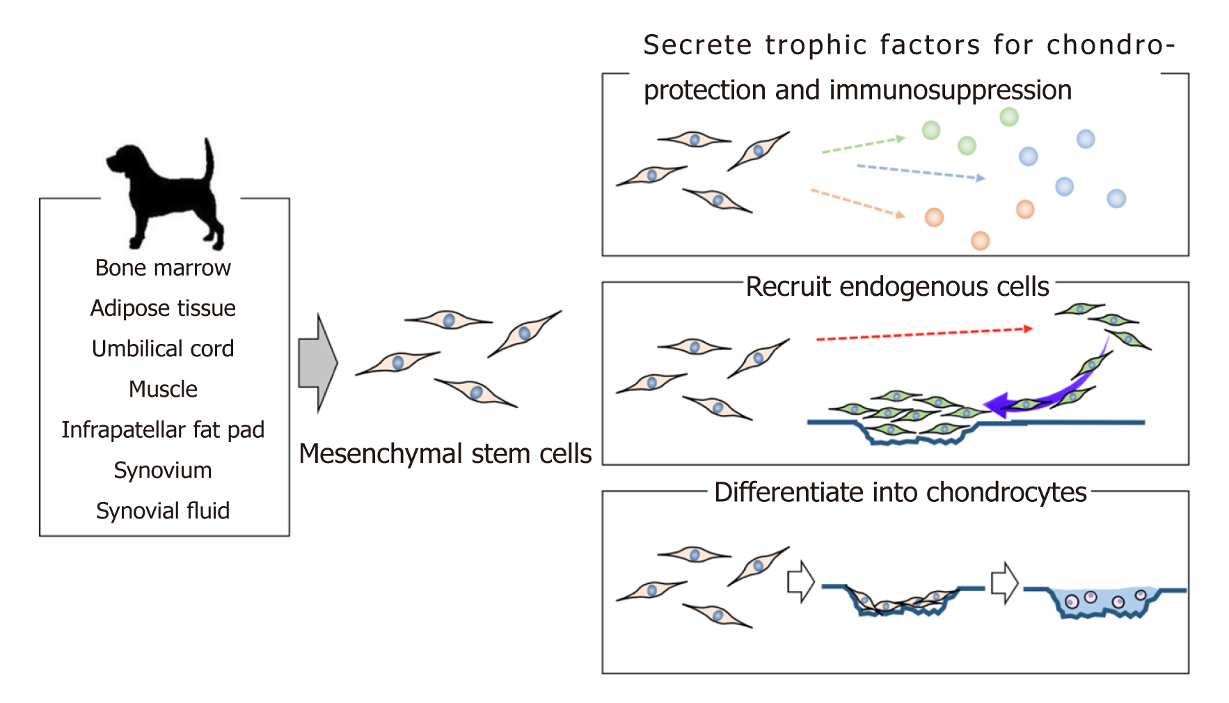
Figure 1 Schematic representation of the role of mesenchymal stem cells for cartilage regeneration.
Mesenchymal stem cells (MSCs) can be isolated from various types of tissues in dogs. MSCs are an attractive therapeutic tool for cartilage regeneration as they can secrete trophic factors for chondroprotection and immunosuppression, recruit endogenous cells to the damaged lesion, and differentiate into chondrocytes, thereby ameliorating the cartilage injury.
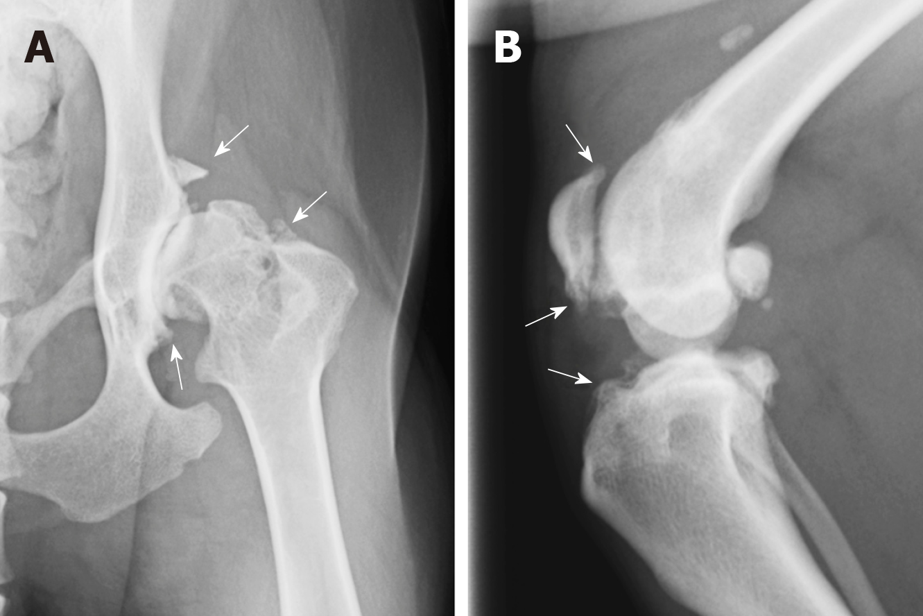
Figure 2 Radiographic findings of osteoarthritis in dogs.
A: A radiographic image of the hip joint in a 8 years old Labrador retriever suffering from osteoarthritis (OA) which results from hip dysplasia; B: A radiographic image of the stifle joint in a 4 years old Shiba suffering from OA which results from cranial cruciate ligament rupture. White arrow: Osteophytes.
Figure 3 Trilineage differentiation of canine mesenchymal stem cells.
A: Adipogenic differentiation confirmed by oil red-o staining. Bar = 50 µm; B: Osteogenic differentiation confirmed by alizarin red. Bar = 100 µm; C: Chondrogenic differentiation confirmed by toluidine blue staining. Bar = 250 µm.
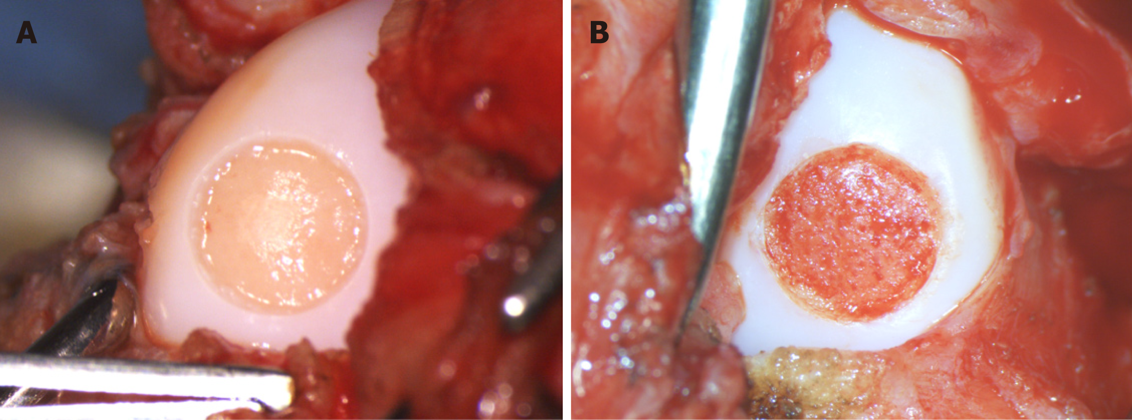
Figure 4 Cartilage defect models in dog stifle joints.
A: A macroscopic image of a partial-thickness cartilage defect model in a dog stifle joint; B: A macroscopic image of a full-thickness cartilage defect model in a dog stifle joint.
- Citation: Sasaki A, Mizuno M, Mochizuki M, Sekiya I. Mesenchymal stem cells for cartilage regeneration in dogs. World J Stem Cells 2019; 11(5): 254-269
- URL: https://www.wjgnet.com/1948-0210/full/v11/i5/254.htm
- DOI: https://dx.doi.org/10.4252/wjsc.v11.i5.254


