Copyright
©The Author(s) 2019.
World J Stem Cells. Dec 26, 2019; 11(12): 1104-1114
Published online Dec 26, 2019. doi: 10.4252/wjsc.v11.i12.1104
Published online Dec 26, 2019. doi: 10.4252/wjsc.v11.i12.1104
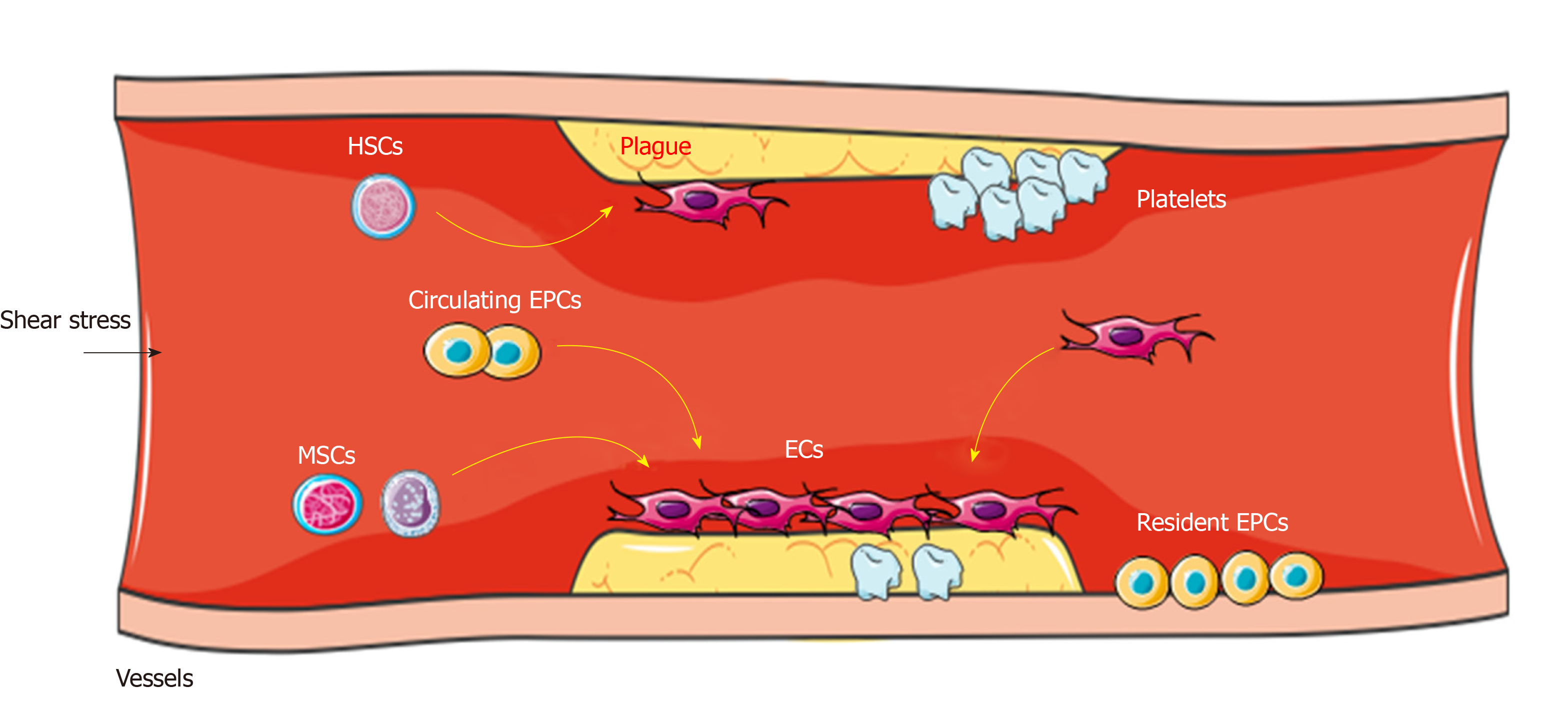
Figure 1 In pathological status, several types of stem cells contribute to the process of vascular repair.
Stem cells such as hemopoietic stem cells, MSCs, circulating EPCs (derived from bone marrow), and resident EPCs are able to sense the activation signals (such as altered shear stress, acute ischemic, and anoxic injury). Then they move to the sites of lesion, differentiate into ECs, produce paracrine signals such as VEGF, PDGF, and SCDF; these actions take part in the vessel regeneration. Importantly, the resident ECs can be activated to proliferation types with a high turnover rate. HSCs: Hemopoietic stem cells; MSCs: Mesenchymal stem cells; EPCs: Endothelial stem cells; ECs: Endothelial cells; VEGF: Vascular endothelial growth factor; PDGF: Platelet-derived growth factor; SCDF: Stromal cell-derived factor.
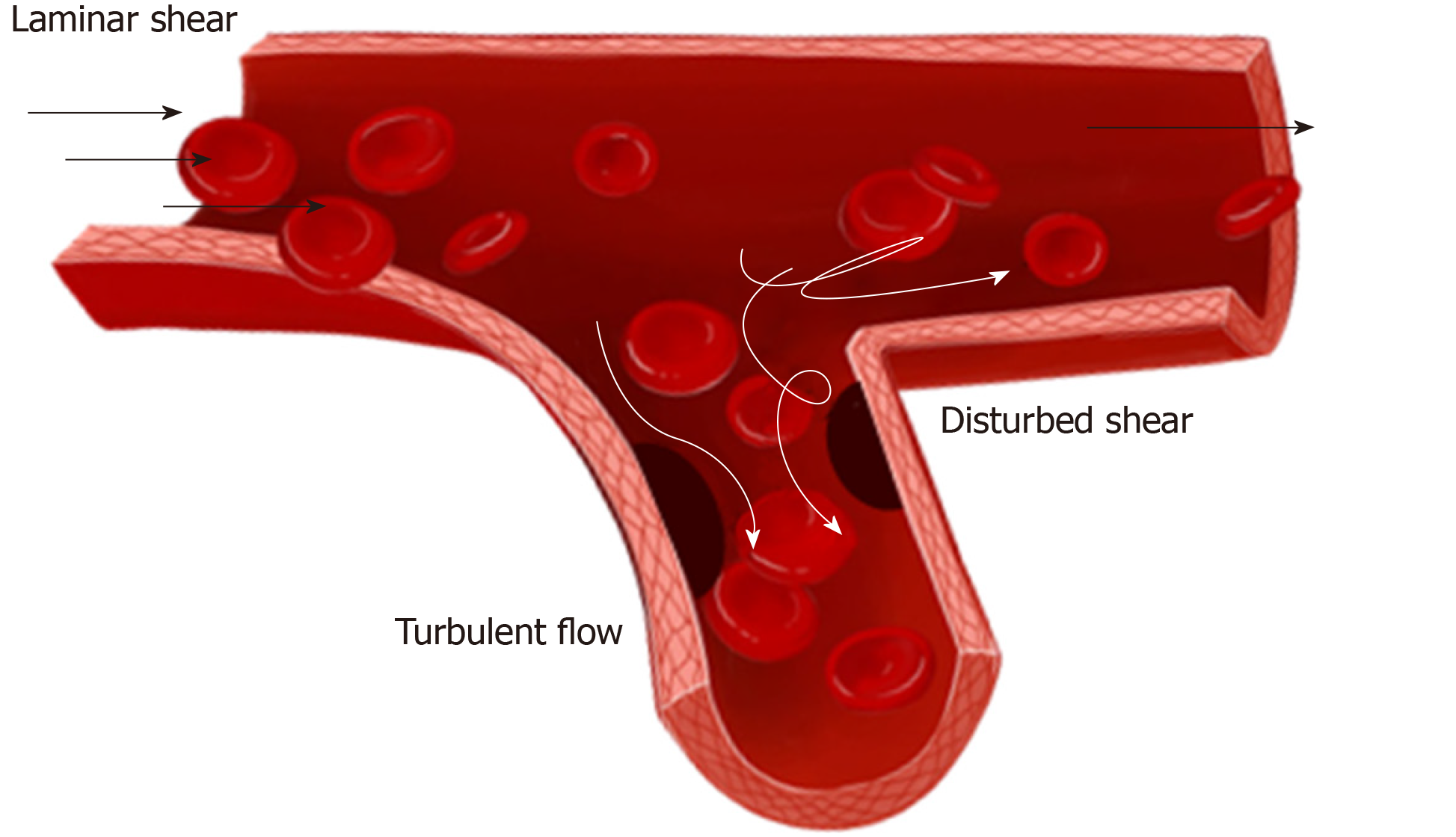
Figure 2 Different patterns of blood flow in vessels.
Arterial flow patterns are exquisitely sensitive to vessel geometry including branching, bifurcation, and curvature. The ECs are exposed to turbulent flow or disturbed shear in the branch points where the atherosclerosis easily developed.
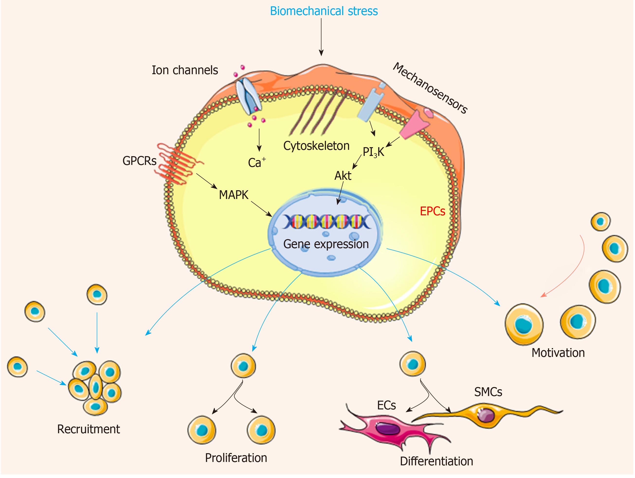
Figure 3 Biomechanical stress stimulates the mechanosensitive molecules on the EPC surface, and then induces cytoskeleton rearrangement and activates series of downstream signaling pathway.
The following cascades lead to change of gene expression, ultimately influencing the cell fundamental activities including cell proliferation, differentiation, recruitment, and motivation, depending upon the need of vessel repair. EPCs: Endothelial progenitor cells; ECs: Endothelial cells; SMCs: Smooth muscle cells. The black arrow points out the direction of stress.
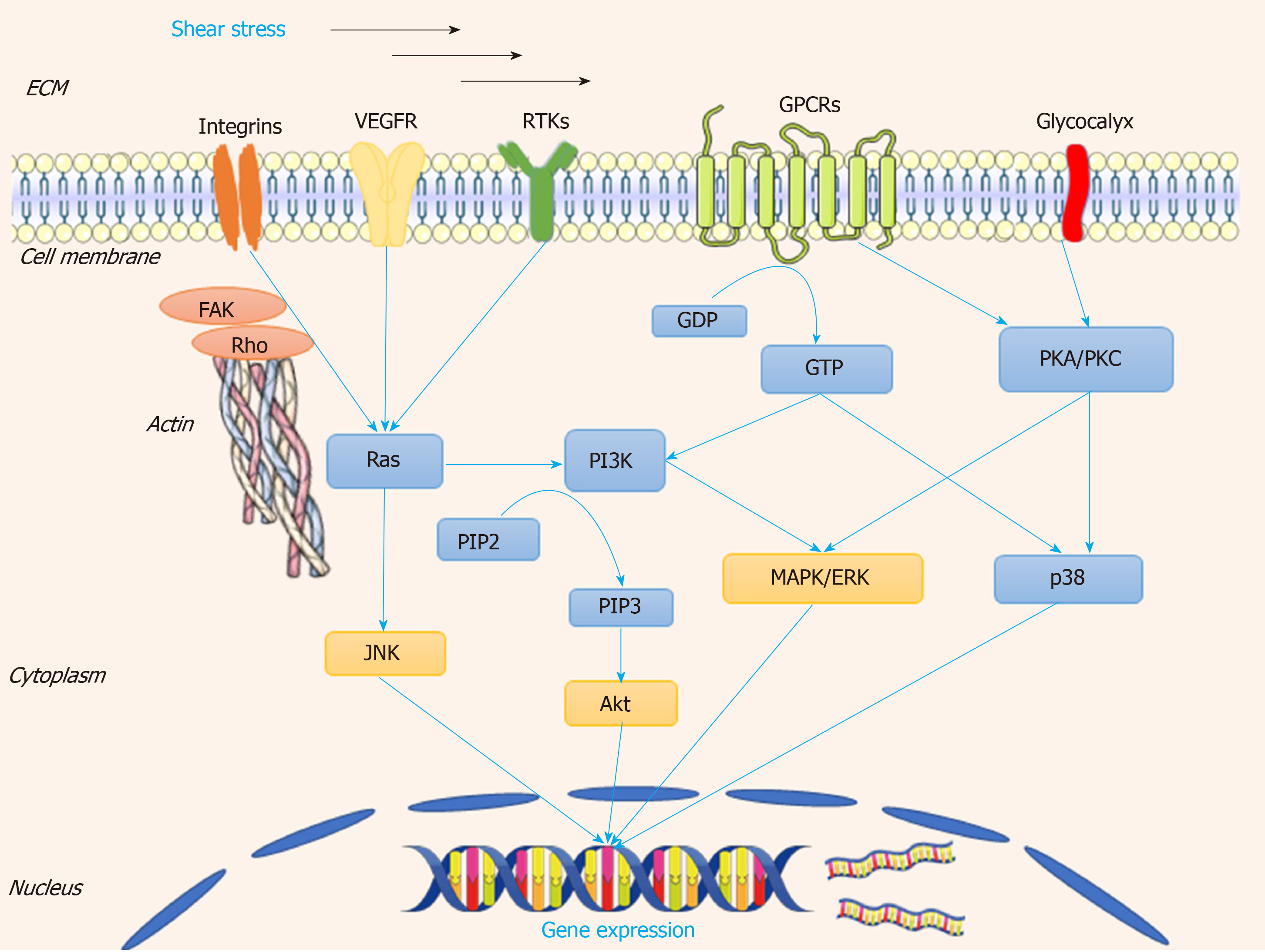
Figure 4 Transduction patterns of mechanical signals into the biological signals, when the shear stress is applied to the cellular surface.
This figure describes how the mechanosensitive molecules such as integrins, VEGFR, RTKs, and GPCRs sense the shear stress, induce cytoskeleton reorganization, and activate various signaling pathways including Ras-PI3K-Akt, Ras-JNK, and PKC/PKA-MAPK-Akt. These signaling cascades influence the gene expression of EPC, ultimately regulating vessel maintenance and reformation. VEGFR: Vascular endothelial growth factor receptor; GPCR: G protein-coupled receptor; RTKs: Receptor tyrosine kinases; PKC: Protein kinase C; MAPK: Mitogen-activated protein kinase; ERK: Extracellular signal-regulated kinase.
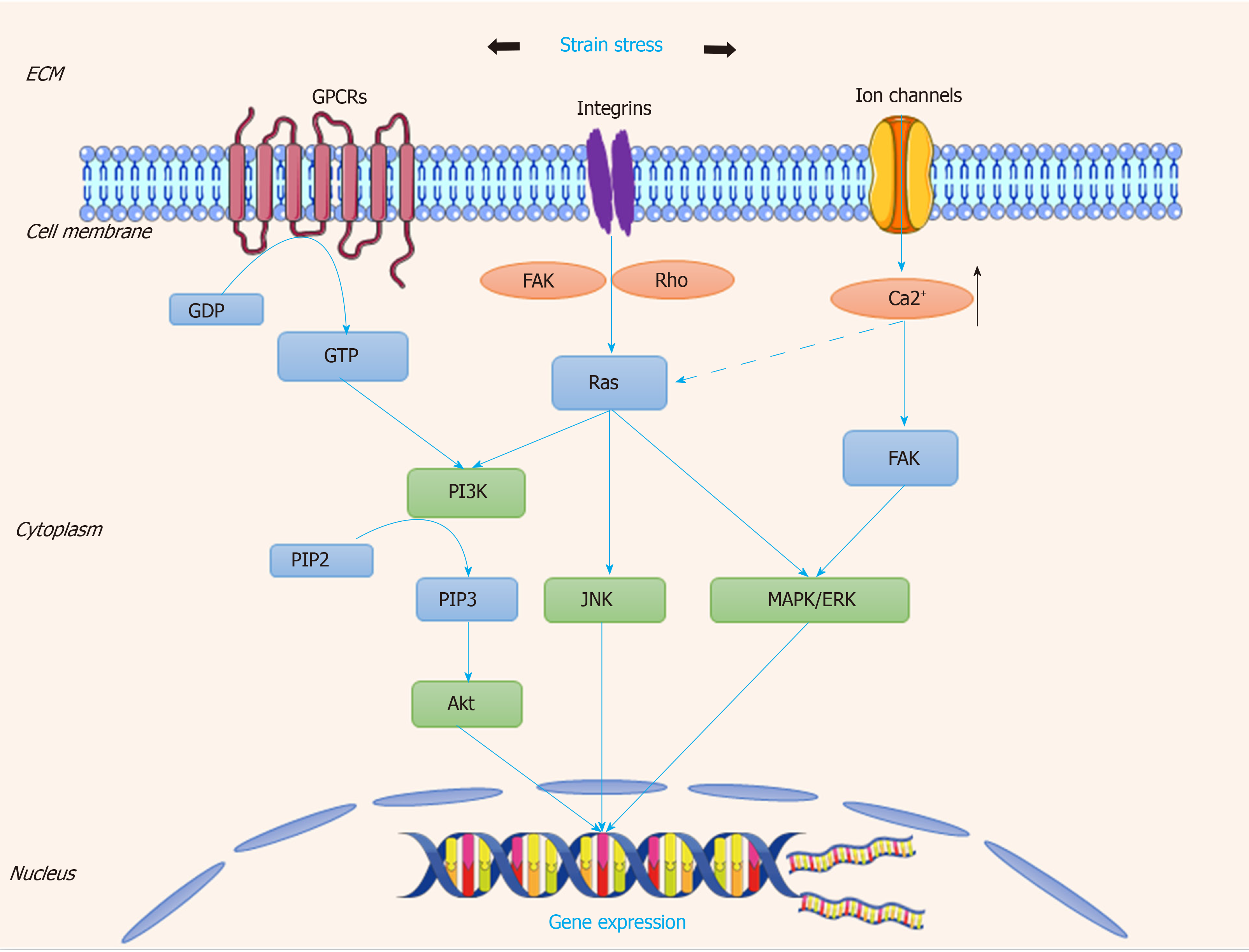
Figure 5 Transduction patterns of mechanical signals into the biological signals, when the strain stress is applied onto the cellular surface.
This figure describes how the mechanosensitive molecules such as integrins, GPCRs, and ion channels sense the strain stress and trigger downstream signaling cascades, then initiate transcription and translation of various genes for cell events in the process of vessel repair. GPCR: G protein-coupled receptor; RTKs: Receptor tyrosine kinases; PKC: Protein kinase C; MAPK: Mitogen-activated protein kinase; ERK: Extracellular signal-regulated kinase.
- Citation: Tian GE, Zhou JT, Liu XJ, Huang YC. Mechanoresponse of stem cells for vascular repair. World J Stem Cells 2019; 11(12): 1104-1114
- URL: https://www.wjgnet.com/1948-0210/full/v11/i12/1104.htm
- DOI: https://dx.doi.org/10.4252/wjsc.v11.i12.1104


