Published online Oct 15, 1998. doi: 10.3748/wjg.v4.i5.409
Revised: July 20, 1998
Accepted: August 16, 1998
Published online: October 15, 1998
AIM: To establish a liver metastasis model of human colorectal carcinoma in nude mice. METHODS: Orthotopic transplantation of histologically intact colorectal tissues from patients into colorectal mucosa of nude mice. Tumorgenicity, invasion, metastasis and morphological characteristics of the transplanted tumors were studied by light microscopy, electron microscopy and immunohistochemistry.
RESULTS: Liver metastasis models of human colon carcinoma (HCA-HMN-1) and human rectal carcinoma (HRA-HMN-2) were established after sceening from 34 colorectal carcinomas.They had been passaged in vivo for 18 and 21 generations respectively. There were lymphatic, hemotogenous and implanting metastasesis. CEA secretion was maintained after transplantation. The primary and liver metastatic tumors were similar to the original human carcinoma in histopathological and ultrastructural features, DNA content and chromosomal karyotype.
CONCLUSION: The liver metastasis models provide useful tools for the study of mechanism of metastasis and its treatment of human colorectal cancer.
- Citation: Liu QZ, Tuo CW, Wang B, Wu BQ, Zhang YH. Liver metastasis models of human colorectal carcinoma established in nude mice by orthotopic transplantation and their biologic characteristic. World J Gastroenterol 1998; 4(5): 409-411
- URL: https://www.wjgnet.com/1007-9327/full/v4/i5/409.htm
- DOI: https://dx.doi.org/10.3748/wjg.v4.i5.409
Some models of nude mice that fresh human colorectal carcinoma tissue or cells were successfully transplanted subcuteneously have been reported at home and abroad[1,2]. But until now there has been no report on a liver metastasis model of human colorectal carcinoma established by orthotopic transplantation in nude mice in China. Based on our previous models of human liver and pancreas carcinoma by orthotopic transplantation[3,4], we established liver metastasis models of colon and rectum carcinoma with a spontaneous metastasis rate of 100%.
BALB/C-nu/nu nude mice were provided by Institute of Oncology, Chinese Academy of Medical Sciences. aged 3-5 weeks, weighing 17 g-20 g, fed under the SPF (specific-pathogen-free) enviroment.
Specimens were obtained from resections of patients with colorectal carcinoma (16 cases of poorly differentiated infiltrating colon adenocarcinoma and 14 cases of poorly differentiated infiltrating rectum adenocarcinoma). The tissues were cut by aseptic manipulation and put into culture liquid RPMI 1640 and cut into pieces of 1 mm × 1 mm × 1 mm for transplantation.
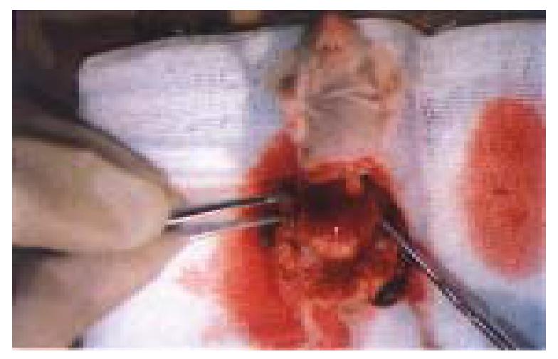
Nude mice were anaesthetized with pentobarbital injection via peritoneal cavity and through the middle incision the colon or rectum were identified. Two tissue masses were sewn into the submucosa from outside the lumen with 0/10 no-injury thread. The growth of the tumor was observed every day. One nude mice who was about to die was killed by cervical vertebra dislocation and the tumor tissues were taken out. A part of tumor tissue was transplanted in situ to other nude mice (5-16 nude mice once) and passaged by the method of primary-generation transplantation continously the remaining part was frozen in liquid nitrogen. Other animals were reserved to observe the tumor metastasis.
Autopsy and histology All the nude mice were examined carefully, the volume of the tumor in situ, and the liver or the other organs and all the abdominal lymph nodes were detected. Specimens were fixed in 10% formalin and hemotoxylineosin stained. Some sections underwent CEA histochemistry study (ABC procedure). The whole liver was insected into 10-20 pieces of slices at different levels to observe the liver metastasis focus.
Tumor in situ and liver metastatic carcinoma were double fixed in 2.5% glutaraldehyde and 1% osmium acid and embraced in Epon-812. After ultramicrotomy, the section was uranium-lead double stained and observed under the TEM (Philips-CM10).
CEA and CA-50 in serum and tumor tissues in situ were measured by radio-immunologic method.
Human tumors, tumor in situ, liver metastatic carcinoma of the 1st, 5th, 10th and 15th generation were analyzed according to the method of reference 5.
Tumor in situ and metastatic liver carcinoma were studied following the methods of Deaven’s and Peterson’s.
After 34 specimens of fresh human colo-rectal carcinoma were orthotopic transplanted to the nude mice, 21 cases were successful one strain of liver metastatic carcinoma model from colon carcinoma was screened, which is named HCA-HMN-1 and another strain of liver metastatic carcinoma model from rectum carcinoma is named HRA-HMN-2.
HCA-HMN-1 was derived from a 37-year-old male, patient with poorly differentiated infiltrating adenocarcinoma accompanied with the metastasis of lymph nodes. This tumor tissue was transplanted orthotopically to 5 nude mice and had passaged the 18th generation. The mean latent period was 10 days. A total of 102 nude mice received the grafting and 30 days fro a generation. Both passaging survival rate and resuscitation rate were 100% (17/17). Necroscopy found that transplanted tumor grew locally infiltrating in large scale. Liver metastatic tumor, abdominal lymph node metastasis and peritonealcavity metastasis could be seen. Pathological evidence suggested that 102 nude mice had liver and lymph node metastasis and 27 had metastasic lung carcinoma.
HRA-HMN-2 was derived from the tumor of a 41-year-old female, patient with poorly differentiated infiltrating adenocarcinoma complicated with the lymph node and liver metastasis. The primary tumor tissue and metastatic liver tumor were transplanted to 8 nude mice successfully and have passaged the 21st generation. The mean latent period was 7 days, and 20 days for one generation. A total of 126 nude mice received the grafting. Both passaging survival rate and resuscitation rate were 100% (17/17). Necroscopy found that both the primary tumor in situ and liver metastatic tumor by orthotopic transplantation grew intensively in mucosa of the rectum in situ accompanying the metastasis of liver and local lymph nodes. The nude mice transplanted with liver metastatic tumor had bloody ascites and extensive tumor seeding in peritoneum. Pathological evidence suggested that the 126 nude mice all had liver and lymph node metastasis, and 31 had lung metastatic and 23 ovarian metastasis.
Liver metastasis appeared at the end of 3 weeks after transplantation. The most of metastatic foci were located in the right lobe of the liver, which were of mononoeud type. A few had polynoeud in both right and left lobes. The diameter of tumor ranged from 0.4 mm to 2.5 cm. Some liver tissues of nude mice could be replaced by metastasic tumor (Figure 1). Pathohistological evidence documented that liver metastatic carcinoma was poorly differentiated colorectal adenocarcinoma.
Lymph node metastasis was limited in the abdominal cavity. Metastasis of lymph nodes of colon, retum, inguinal, mesentery and a few hilur hepatis could be seen, which can be devided into three stages: in early stage, carcinoma cell mass only appeared in afferent lymphatic and marginal sinus; in middle stage, cancer cell progressively invated the paracortical zone and medulla; and in late stage, the whole lymph node was nearly occupied by the cancer cells except for the residual margins.
Under light microscopy, the distribution of HCA-HMN-1 cancer cells was characterized by blocks and mass with small adenoid structure which had plenty of plasma, irregular giant nuclear, obvious nucleoli and polynuclear tumor giant cells. The HRA-HMN-2 cells appeared circle and ellipsoid in shape with, abundant plasma, heavyly stained giant nuclear, obvious nucleoli, rough and large karyotin granulae, and tumor giant and pomynuclear tumor cells appeared frequently. TEM showed that HCA-HMN-1 cancer cells had irregular shape, heteromorphic giant nucleus and polynucleoli. The heterochromatin was distributed around the uncleus. In cytoplasma there were mitochondria and endoplasmic reticulum, occassionally microvilli. Intracellular conjunction was complex or desmosome. Liver metastatic tumors showed the typical ultrastructure of the poorly differentiated colon adenocarcinoma. HRA-HMN-2 cells were circled as adenoid lumen in which the surface had a little microvilli in which the direction, length and diameter were highly different. Permutation of peripheral cancer cells was irregular, with large nucleus, obvious karyotin and nucleoli, and wrapped nuclear membrane. In cytoplasma, there was a moderate quantity of rough endoplasmic reticulum and a small amount of edema mitochondria. The maldevelopment desmosome-like intracellular conjunction was seen in a few cells. Liver metastatic tumor presented the ultrastructural features of poorly differentiated rectal adenocarcinoma.
The mean value of the CEA in tumor tissues was 79.8 mg/L, and CA-50 was 57.6 U/mL; and in peripheral blood, 24.3 mg/L and 33.6 U/mL respectively.
Whole-plasma-positive cells could be seen in the transplanted tumor cells and liver metastatic carcinoma cells of nude mice.
The DNA ploidy of the original human tumor specimen, implanted tumor of nude mice and liver metastatic tumor tissue were all nearly tetraploid.
Transplanted tumor and liver metastatic carcinoma of nude mice had the similar karyotype to human tumor cells. The number of chronsome ranged from 77 to 86, which demoustrcted as supertriploid or subtetraploid.
Liver is the commonest metastatic locus of colo-rectal carcinoma[6]. The incidence of liver metastasis might be 20%-40% when the diagnosis of colo-rectal carcinoma was documented and 40%-50% after radical resection of colo-rectal carcinoma. About 67% of the patients who died of metastasis had liver metastasis carcinoma[7]. The deep invasion and metastasis did not occur in the subcutaneous tumor models by implanting fresh human colorectal carcinoma tissue or cells into nude mice[1,2]. The liver metastatic tumor produced by endosplenic grafting of colon carcinoma cell line was experimental but not spontaneous[8], and was not ideal model to be used for studying the metastatic liver carcinoma.
To study the mechanism and therapy of liver metastasis, a model of ortotopic transplantation, which was similar to human being and non-cell-line, should be established. In this study two strains of models, called HCA-HMN-1 and HRA-HMN-2 were established, which perfectly demonstrated the clinical metastatic process of human colo-rectal carcinoma not only in the aspect of the invasive growth and tumor formation in the colo-rectum of nude mice but also in keeping 100% of the liver and lymph node metastatic rate. So our models subjectly simulated the clinical features of patients with liver metastasis.
The results showed that in colon carcinoma the two models, after they continuously passaged for two years, the feature of chronsome and histological ultrastructure were totally the same as that of the original tumors which can secrete the CEA and CA-50. That liquid nitrogen frozen tissue of transplanted tumor can still be reimplanted successfully suggested that the models had stable biologic features.
There were two causes of liver metastasis. Tumor cells itself had strong potential ability of invasion. This study successfully used two specimens of Duck’s D stage of colorectal carcinoma; The microcirculation (the colo rectum has special structures, and plenty of lymph and blood supply. It is the colo-rectal microcirculation of nude mice which was so similar to the that of human being that the transplanted tumor of nude mice has the similar clinical process of liver metastasis from human colo-rectal carcinoma. We consider that only the orthotopic transplantation method of nude mice can produce metastasis similar to patients with higher spontaneous metastasis rate than that of other models.
Our results show that HCA-HMN-1 and HRA-HMN-2, have obtained the biologic features in aspects of morphology, biochemistry, immunology and genetics, and are ideal models to study the mechanism of human colo-rectal carcinoma metastasis and anti-metastasis therapy.
Project supported by the Military Science Foundation, No.970056.
| 1. | Rygaard J, Povlsen CO. Heterotransplantation of a human malignant tumour to "Nude" mice. Acta Pathol Microbiol Scand. 1969;77:758-760. [PubMed] [DOI] [Full Text] |
| 2. | Wu BQ, Sun YK, Zhen J. The study on the transplanted tumor of human mucus adenoicarcinoma in nude mice and its biologic characteristics. J B Med U. 1982;14:205-208. |
| 3. | Tuo CW, Liu QZ, Wu BQ. The models of human liver cancer transplanted orthotopicly into nude mice and its biologic characteristics. Chin J Experim Surg. 1992;9:28-29. |
| 4. | Liu QZ, Tuo CW, Wu BQ. The models of human pancreas cancer transplanted orthotopicly into nude mice and its biologic characteristics. Chin J Oncol. 1992;14:403-406. |
| 5. | Vindeløv LL, Christensen IJ, Nissen NI. A detergent-trypsin method for the preparation of nuclei for flow cytometric DNA analysis. Cytometry. 1983;3:323-327. [PubMed] [DOI] [Full Text] |
| 6. | Nordlinger B, Panis Y, Puts JP, Herve JP, Delelo R, Ballet F. Experimental model of colon cancer: recurrences after surgery alone or associated with intraperitoneal 5-fluorouracil chemotherapy. Dis Colon Rectum. 1991;34:658-663. [PubMed] [DOI] [Full Text] |
| 7. | Cady B, Stone MD. The role of surgical resection of liver metastases in colorectal carcinoma. Semin Oncol. 1991;18:399-406. [PubMed] |
| 8. | Morikawa K, Walker SM, Jessup JM, Fidler IJ. In vivo selection of highly metastatic cells from surgical specimens of different primary human colon carcinomas implanted into nude mice. Cancer Res. 1988;48:1943-1948. [PubMed] |


