Copyright
©The Author(s) 2018.
World J Gastroenterol. Jan 21, 2018; 24(3): 360-370
Published online Jan 21, 2018. doi: 10.3748/wjg.v24.i3.360
Published online Jan 21, 2018. doi: 10.3748/wjg.v24.i3.360
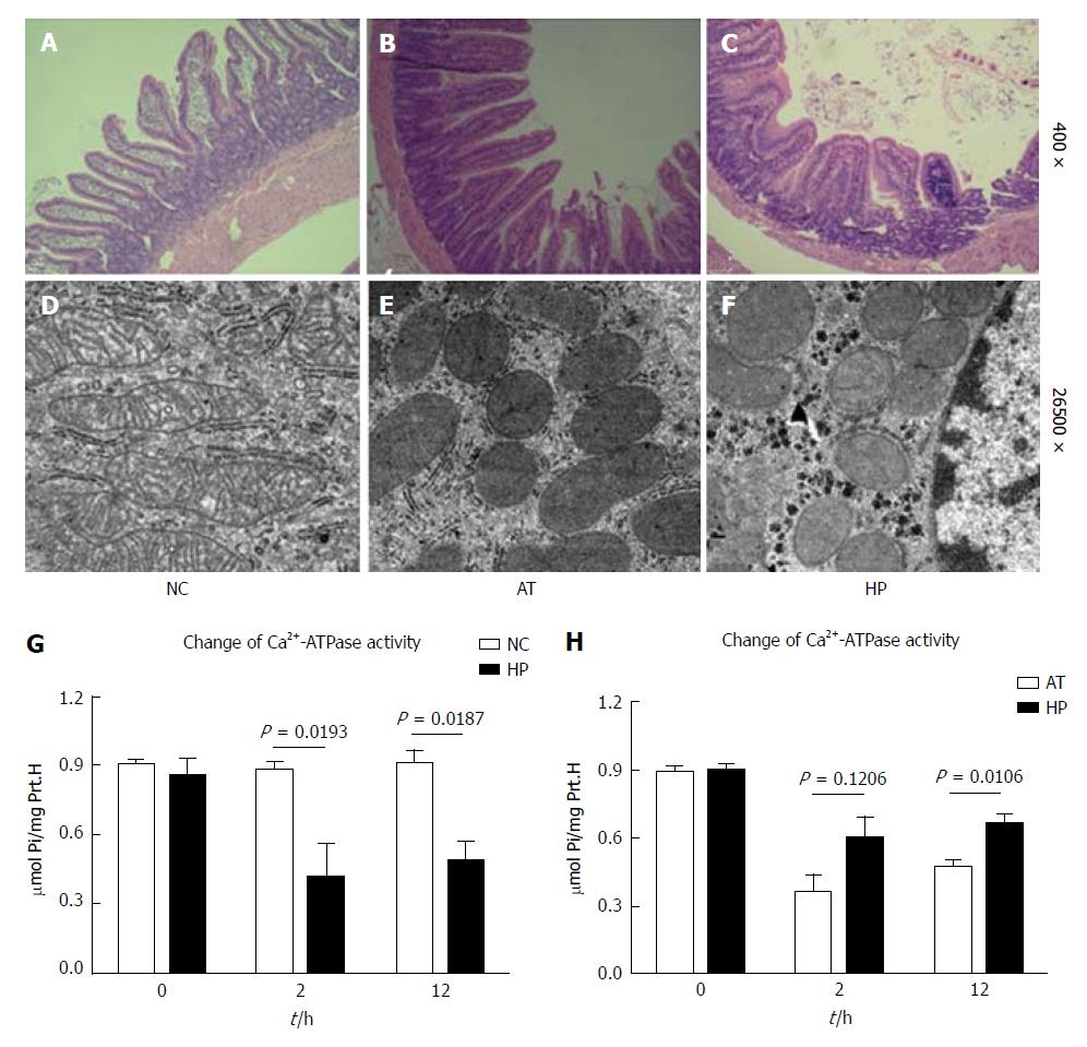
Figure 2 Hypoxia-induced HIF-1α expression increases intestinal cellular mitochondrial integrity and Ca2+-ATPase activity after I/R injury.
A-C: Pathological changes in intestinal cells are shown (HE, 10 × 20). A: The normal morphology of intestinal cells in the NC group; B: The intestinal cells in the AT group exhibited oedema and red blood cell deposition and blood clots in capillary vessels; the villus epithelial cells were shedding, had severely damaged glands, and had been infiltrated by inflammatory cells; C: The intestinal cells in the HP group exhibited slight oedema and fewer inflammatory cells. The villus epithelial cells were less damaged. D-F: The mitochondrial changes in intestinal cells are shown (× 46000). D: The normal morphology of mitochondria in the NC group; E: The mitochondria in the AT group appeared swollen, round, and degenerated. The visible mitochondrial cristae appeared less fractured or disappeared; F: The mitochondria in the HP group showed less swelling and fewer cristae. The endoplasmic reticulum structure had survived; G: The Ca2+-ATPase activity in the AT group was significantly lower compared with the NC group, which decreased to the lowest level at postoperative 2 h (AT group vs NC group: P < 0.05) and then recovered; H: The Ca2+-ATPase activity in the HP group was lower after the operation but was significantly higher than that in the AT group at postoperative 2 h (HP group vs AT group: P < 0.05).
- Citation: Ji ZP, Li YX, Shi BX, Zhuang ZN, Yang JY, Guo S, Xu XZ, Xu KS, Li HL. Hypoxia preconditioning protects Ca2+-ATPase activation of intestinal mucosal cells against R/I injury in a rat liver transplantation model. World J Gastroenterol 2018; 24(3): 360-370
- URL: https://www.wjgnet.com/1007-9327/full/v24/i3/360.htm
- DOI: https://dx.doi.org/10.3748/wjg.v24.i3.360


