Minireviews Open Access
Copyright ©The Author(s) 2016. Published by Baishideng Publishing Group Inc. All rights reserved.
World J Gastroenterol. Mar 7, 2016; 22(9): 2760-2770
Published online Mar 7, 2016. doi: 10.3748/wjg.v22.i9.2760
Life and death at the mucosal-luminal interface: New perspectives on human intestinal ischemia-reperfusion
Joep Grootjans, Kaatje Lenaerts, Cornelis HC Dejong, Joep PM Derikx, Department of Surgery, NUTRIM School of Nutrition and Translational Research in Metabolism, Maastricht University Medical Center, 6200 MD Maastricht, The Netherlands
Joep Grootjans, Brigham and Women’s Hospital, Harvard Medical School, Boston, MA 02115, United States
Wim A Buurman, NUTRIM School of Nutrition and Translational Research in Metabolism, Maastricht University Medical Center, 6200 MD Maastricht, The Netherlands
Author contributions: Grootjans J and Derikx JPM wrote the manuscript; Grootjans J, Lenaerts K, Buurman WA, Dejong CHC and Derikx JPM provided important intellectual contribution to the manuscript.
Supported by Dutch Gastroenterology and Hepatology Society (MLDS grant WO10-57 to Dejong CHC and Lenaerts K); Career Development Grant CDG (to Derikx JPM); and The Netherlands Organisation for Scientific Research (Rubicon grant 825.13.012 to Grootjans J).
Conflict-of-interest statement: No conflicts of interest exist.
Open-Access: This article is an open-access article which was selected by an in-house editor and fully peer-reviewed by external reviewers. It is distributed in accordance with the Creative Commons Attribution Non Commercial (CC BY-NC 4.0) license, which permits others to distribute, remix, adapt, build upon this work non-commercially, and license their derivative works on different terms, provided the original work is properly cited and the use is non-commercial. See: http://creativecommons.org/licenses/by-nc/4.0/
Correspondence to: Joep Grootjans, MD, PhD, Department of Surgery, NUTRIM School of Nutrition and Translational Research in Metabolism, Maastricht University Medical Center, 6200 Maastricht, The Netherlands. j.grootjans@maastrichtuniversity.nl
Telephone: +31-43-3881497 Fax: +31-43-3884154
Received: June 22, 2015
Peer-review started: June 25, 2015
First decision: July 14, 2015
Revised: September 24, 2015
Accepted: December 14, 2015
Article in press: December 14, 2015
Published online: March 7, 2016
Processing time: 254 Days and 9.8 Hours

Abstract

Intestinal ischemia is a frequently observed phenomenon. Morbidity and mortality rates are extraordinarily high and did not improve over the past decades. This is in part attributable to limited knowledge on the pathophysiology of intestinal ischemia-reperfusion (IR) in man, the paucity in preventive and/or therapeutic options and the lack of early diagnostic markers for intestinal ischemia. To improve our knowledge and solve clinically important questions regarding intestinal IR, we developed a human experimental intestinal IR model. With this model, we were able to gain insight into the mechanisms that allow the human gut to withstand short periods of IR without the development of severe inflammatory responses. The purpose of this review is to overview the most relevant recent advances in our understanding of the pathophysiology of human intestinal IR, as well as the (potential) future clinical implications.

Key Words: Small intestinal ischemia; Colon ischemia; Epithelial lining; Ischemia-reperfusion; Human models

Core tip: To improve our knowledge on the pathophysiology of human intestinal ischemia-reperfusion (IR), a new human experimental model was developed in recent years. In the current review, we overview and discuss the most important new insights on intestinal IR in man.


INTRODUCTION

Intestinal ischemia-reperfusion (IR) is a frequently observed phenomenon in health and disease[1,2]. It occurs as part of normal physiology, for example in exercising healthy individuals[3,4], in whom redistribution of blood flow to the vital organs and muscles leads to significant intestinal hypoperfusion with up to 80% reduced mesenteric blood flow[3,5]. Moreover, intestinal IR occurs in a variety of pathophysiological situations. It is observed in patients with acute mesenteric ischemia due to mesenteric arterial embolism or venous thrombosis. In addition, intestinal IR occurs in situations with severe blood loss and/or hypovolemia, leading to redistribution of blood flow to the most important organs, including heart and brains. This form of intestinal IR is frequently observed in patients undergoing major surgery or in patients with trauma, shock or sepsis[1,6]. Lastly, intestinal ischemia is a well-known player in the development and perpetuation of intestinal inflammation, including in patients with inflammatory bowel disease[7,8].

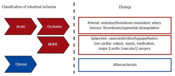
Based on etiological background, intestinal ischemia is divided into a chronic and an acute form (Figure 1). Chronic gastrointestinal ischemia (CGI) is most commonly due to atherosclerotic disease and has been reviewed elsewhere[9,10]. In contrast to CGI, acute intestinal ischemia is a consequence of a rapid reduction in intestinal blood flow, caused by occlusion (occlusive disease) or severe hypoperfusion [non-occlusive mesenteric ischemia (NOMI)] of mesenteric vessels (Figure 1)[1,11,12]. Acute intestinal ischemia is a potentially fatal clinical emergency with an overall mortality of 60% to 80%[1,2,11]. Mortality rates are especially high when ischemia progresses towards bowel necrosis, leading to a severe inflammatory response, sepsis and shock[11,12].

Figure 1
Figure 1 Schematic overview of intestinal ischemia based on etiological background. NOMI: Nonocclusive mesenteric ischemia.

It is remarkable that although medical practice has evolved considerably, the mortality rates of intestinal ischemia did not improve over the past 70 years[1,13]. This is partly a consequence of the continued difficulty to recognize acute mesenteric ischemia at an early stage, which is the key to reducing high morbidity and mortality[14]. The second major reason for intestinal IR-related high morbidity and mortality rates is the paucity in preventive and/or therapeutic options. Obviously, rapid reperfusion after intestinal ischemia is of major importance, but reperfusion can paradoxically also contribute to tissue injury and severe inflammation[11,15,16]. Improved insight into the mechanisms underlying intestinal IR-related complications in man are imperative to work towards novel therapeutic strategies. This review provides an overview of the current knowledge on the pathophysiology of human intestinal ischemia-reperfusion. Development of unique human IR models allowed detailed investigation of the consequences of human intestinal IR on several important players of the intestinal barrier function.

THE HUMAN INTESTINAL EPITHELIAL BARRIER

The primary functions of the human gastrointestinal tract are digestion of food, absorption of nutrients and water, and electrolyte exchange. In addition, the gut provides a barrier between the internal milieu and the potentially toxic intraluminal content, including commensal and pathogenic microorganisms and their products[17,18]. For this purpose, the intestinal tract is equipped with several barriers. The first anatomical site at which the host encounters the gut microbiota is the mucus layer[19,20]. This layer serves as a physical and chemical barrier, preventing adherence of microbiota to the epithelium and impedes translocation of potential pathogens and their toxins to the internal milieu[19-21]. The secreted mucins are continuously produced by specialized goblet cells that are found in the small and large intestinal epithelium. Secretory mucins are stored in bulky apical granules of the goblet cells[21]. The mucin-containing granules are secreted from the apical surface both constitutively and in response to a variety of external stimuli[19,21]. The second line of defence is formed by the intestinal epithelium[22] which consists of a cohesive monolayer of columnar epithelial cells[23], firmly connected by tight junction complexes that seal the paracellular pathway[18,24,25]. Next to providing a physical barrier, enterocytes actively participate in innate immunity, by acting as immune sensors of microbial pathogens and commensal organisms[17]. The signaling loop that mediates the epithelial response to microorganisms is based on sensing of structural motifs, known as pathogen-associated molecular patterns (PAMPs) by pattern-recognition receptors (PRRs) on epithelial cells[26]. PAMPs are expressed by both commensal and pathogenic microorganisms, and include lipopolysaccharide (LPS), lipoprotein and peptidoglycans[17].

A third line of defense is formed by Paneth cells, highly specialized epithelial cells residing in the crypts of the small intestine in between the stem cells[27-30]. Paneth cells are the main source of antimicrobial proteins, including α-defensins and lysozyme, in the small intestine[28,31,32]. These proteins are stored in granules and released into the crypt lumen both constitutively and in response to bacterial threats[32] to prevent microbial invasion[33,34]. In addition, the antimicrobial proteins disseminate into the mucus layer in order to reinforce the mucosal barrier function[33-35]. Lastly, Paneth cells provide critical stem cell niche factors, including EGF, Wnt3 and Notch[36].

From studies in a variety of animal models we have learned that intestinal IR can severely damage the intestinal epithelial barrier. Our current knowledge on (patho)physiological mechanisms of IR-induced intestinal injury in a variety of animal models has recently been excellently reviewed elsewhere[37]. So far, translation to the human setting was only possible in post-mortem studies or in patients undergoing surgery with resection of necrotic intestinal segments after exposure to extensive periods of ischemia, which only allowed investigation of the end-stage of the disease. To overcome these problems, we have developed in recent years novel human experimental intestinal IR models. These models allowed us to investigate the pathophysiology and underlying mechanisms of human small intestinal IR and colonic IR.

THE HUMAN SMALL INTESTINAL IR MODEL

Development of a standardized experimental human model for small intestinal IR was considered to be of significant advantage in addressing clinical questions. Important issues during development of an in vivo human model were first, that it should be ethically justified, safe, and would allow only minimally invasive experimental procedures. Second, the model should enable investigators to expose a healthy intestinal segment to various periods of ischemia and reperfusion. Third, it should allow collection of both tissue and blood during the experimental protocol at set time points, yielding reproducible and consistent results. This led to the inclusion of patients undergoing abdominal surgery with a Roux-Y or similar small intestinal reconstruction (e.g., during pylorus-preserving pancreatico-duodenectomy, pancreatico-jejunostomy, hepatico-jejunostomy, or Frey procedure). In these patients, a variable length of healthy jejunum is usually resected in continuity with the specimen as part of the standard surgical procedure. Approximately 6 cm of this healthy part of jejunum was used for the experiments; therefore no additional tissue had to be resected for study purposes.

In short, after identification and localization of the major anatomical structures and tumor during surgery, a healthy jejunal segment with a central mesenteric arteriole and venule was isolated by transsection at both ends with a linear cutting stapler. This segment was subjected to various periods of ischemia by placing 2 atraumatic vascular clamps across the mesentery. Meanwhile, surgery proceeded as planned. Removal of the clamps led to reperfusion as confirmed by regaining of normal pink color and restoration of intestinal motility. Tissue was sampled at selective time point using the linear cutting stapler, and blood was sampled by direct puncture of the venule draining the isolated jejunal segment. Arterial blood was sampled from the radial artery line, present in all patients as part of routine intraoperative monitoring. This allowed assessing concentration gradients in plasma across the isolated jejunal tissue. At the end of the experiment, 2 cm of jejunum that remained untreated during surgery was resected and served as internal control tissue. This segment underwent similar surgical handling as the isolated part of jejunum while it was not exposed to IR. A more detailed description of the human intestinal IR model, including a diagram of the model, can be found in reference[38].

In this newly developed human experimental model to study small intestinal IR, the maximum time of reperfusion of the isolated ischemic jejunum is dependent on the surgical procedure, i.e., the moment at which the surgeon decides that the pancreatico- or hepatico-jejunal anastomosis has to be created. Using this model, we obtained insight into the pathophysiology of short an prolonged periods of ischemia followed by reperfusion and we elucidated the key molecular events during intestinal IR in man.

SHORT PERIODS OF SMALL INTESTINAL ISCHEMIA WITH REPERFUSION: MECHANISMS TO PREVENT EPITHELIAL LINING DAMAGE

The human intestine is frequently exposed to short periods of ischemia, both in physiological and pathophysiological situations. Maintenance of the intestinal epithelial lining is of major importance, since loss of this barrier facilitates translocation of potentially harmful intestinal luminal contents towards the circulation, causing a severe inflammatory response. Using the human experimental IR model, we obtained insight into the duration of ischemia that the human jejunum can withstand before extensive barrier damage occurs.

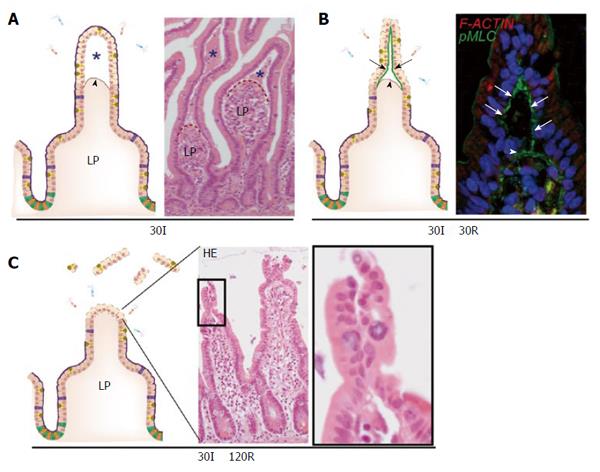
A key feature of short small intestinal ischemia (without reperfusion) is the appearance of subepithelial spaces (Figure 2A). These can be observed as early as 15 min after the onset of ischemia, but are more pronounced at 30 min of ischemia[5,39]. These spaces have been described previously in animal studies and are classically called the spaces of Gruenhagen[40,41]. These subepithelial spaces are the consequence of detachment of the upper, ischemically damaged, epithelial cells from the basal membrane with simultaneous contraction of the basement membrane muscle fibers emanating up to the tips of the villi[6,40]. Detachment of the epithelial cells from their basement membrane was only observed at the villus tips, probably because these cells are more susceptible to ischemia. This is in part due to a countercurrent flow in the villi which causes lower oxygen supply to the upper parts of the villus compared to the crypts[7]. In addition, enterocytes obtain more pro-apoptotic characteristics as they migrate up towards the tip of the villus, where they undergo apoptosis and are shed into the lumen at the end of their life cycle (also called “anoikis”)[42,43]. To our surprise, detachment of epithelial cells at the villus tip did not directly result in a disrupted epithelial lining. Instead, we observed that early after onset of reperfusion, the sheets of loose epithelial cells were pulled together[5,39]. Our studies suggested that this was achieved by accumulation of non-muscle type myosin fibers, phosphorylated myosin light chain (pMLC), at the basal side of the loose epithelial cells leading to active constriction of the IR-damaged cells at the villus tips, resulting in a dramatic reduction in wound size (Figure 2B)[39]. This phenomenon, which we termed zipper-like epithelial constriction, morphologically resembled purse-string contraction in small epithelial wounds, which is similarly characterized by accumulation of actin and myosin at the apical side of leading edge cells followed by active contraction to close small epithelial defects[44-47]. Indeed, accumulation of filamentous actin and pMLC was observed in the epithelial defects at 120 min of reperfusion, indicating that purse string contraction is involved in wound healing of IR-damaged small intestine.

Figure 2
Figure 2 The human small intestine has efficient mechanisms to prevent excessive epithelial lining damage. A: Cartoon and hematoxylin-eosin staining demonstrating the appearance of subepithelial spaces (asterisks) after ischemia as a result of retraction of the basement membrane (arrowhead); B: Early during reperfusion, loose IR-damaged epithelial sheets are pulled together through active contraction of pMLC at the basal side of epithelial cells (green line, arrows), bringing these cells together; C: Zipper-like constriction of the epithelium is associated with rapid restoration of the epithelial lining and prevents exposure of lamina propria to intraluminal content. LP: Lamina propria; pMLC: Phosphorylated myosin light chain; F-ACTIN: Filamentous actin.

The combined mechanisms of villus retraction, constriction of loose epithelial cells and rapid pMLC-mediated closure of the epithelial defects resulted in a morphologically restored epithelial lining within 120 min of reperfusion of ischemically damaged jejunum (Figure 2C). This mechanism could therefore be involved in limiting bacterial translocation and vigorous inflammation in response to environmental conditions that cause a mild decreased splanchnic perfusion, such as physical exercise and surgery[48].

The fact that IR-induced zipper-like constriction of the epithelium was induced even before epithelial cells were shed into the lumen, emphasizes that it is not only essential to close small epithelial defects but importantly, it is crucial to limit the development of epithelial wounds.

PROLONGED SMALL INTESTINAL ISCHEMIA WITH REPERFUSION: PHYSICAL INTESTINAL BARRIER INTEGRITY LOSS AND OCCURRENCE OF LOCAL INFLAMMATION

In contrast to short periods of ischemia of the small intestine, the epithelial lining starts to disintegrate after more than 45 min of small intestinal ischemia. Damage of villus tips further continues during reperfusion, and apoptosis of enterocytes at the villus tips becomes particularly apparent at 30 min of reperfusion. It seems that restorative mechanisms as described above, preventing exposure of the lamina propria to the intraluminal content, fail after longer periods of small intestinal ischemia[49]. This is consistent with data from animal models, in which it has been shown that with increasing duration of the ischemic periods, progressive cell death occurs from villus to crypt[40,50]. Additionally, restoration of IR-induced damage was not achieved after 120 min of reperfusion in tissue exposed to > 45 min of ischemia. This results in prolonged exposure of lamina propria immune cells to PAMPs and damage-associated molecular patterns (DAMPs) which both elicit inflammation.

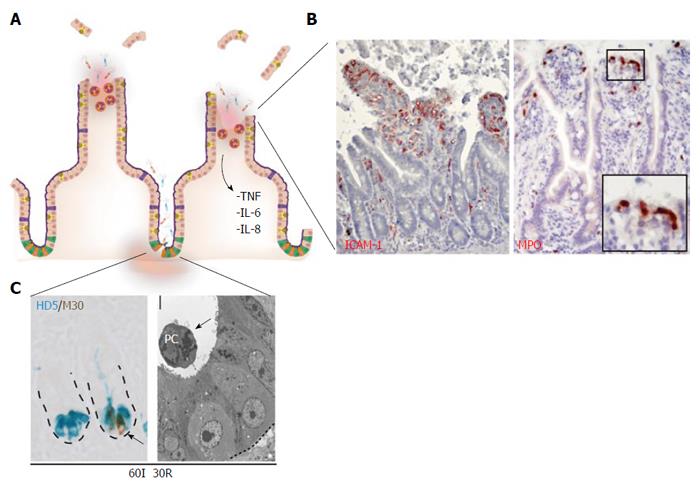
Consequently, and in contrast to the findings presented for short periods of small intestinal ischemia, an inflammatory response develops during the reperfusion phase after prolonged ischemia (Figure 3A). First, enhanced expression of intercellular adhesion molecule-1 (ICAM-1) becomes apparent on the endothelium early during reperfusion, which is accompanied by sequestration and influx of neutrophils into the damaged villus tips (Figure 3B)[49]. Neutrophils accumulated just beneath the damaged epithelial lining, which has been described to play a role in sterilizing the wound (Figure 3B, right panel)[6,51]. However, it should be taken into account that activated neutrophils can also augment to reperfusion-induced damage, since local release of their antimicrobial proteins including myeloperoxidase results in formation of reactive oxygen species (ROS)[52,53]. Second, the inflammatory response was accompanied by complement activation, represented by the deposition of activated C3, a downstream target in the complement activation cascade, in IR-damaged epithelial cells in the lumen[49].

Figure 3
Figure 3 Prolonged small intestinal ischemia-reperfusion results in physical and immunological barrier function loss and inflammation. A: Prolonged IR leads to disruption of the epithelial lining (physical barrier integrity loss) and Paneth cell loss (immunological barrier integrity loss); B: Inflammatory responses are characterized by increased endothelial expression of ICAM-1 (left panel) with sequestration of MPO-positive neutrophils into the villus tips (right panel), and increased expression and release of inflammatory cytokines including IL-6, IL-8 and TNF; C: Left panel: Prolonged IR leads to Paneth cell apoptosis, as shown by the co-localization of M30 (brown: apoptosis) and human defensin 5 (blue: Paneth cells). Right panel: EM picture of apoptotic Paneth cell, shed into the crypt lumen. ICAM-1: Intercellular adhesion molecule-1; MPO: Myeloperoxidase; HD5: Human defensin-5.

Complement activation has been identified as a key mediator of IR-induced inflammation in mainly murine IR models[54], with involvement of the classical[55], the alternative[56,57] as well as the lectin[58] activation pathway. Complement activation fragments have chemoattractant properties and are capable of inducing production of chemokines and cytokines[59]. In line with this, increased mRNA expression of pro-inflammatory cytokines interleukin (IL)-6, IL-8 and tumor necrosis factor (TNF)-α was observed as early as 30 min after induction of reperfusion, which was accompanied by increased arteriovenous concentration differences of IL-6 and IL-8 across the small intestine[49].

PROLONGED SMALL INTESTINAL IR: IMMUNOLOGICAL BARRIER FUNCTION LOSS AND INCREASED SYSTEMIC INFLAMMATION

Apart from the physical barrier, mainly comprised of a mucus layer and an epithelial lining, the intestine is also dependent on its immunological barrier function to prevent bacterial translocation. An important player in the immunological defense is the Paneth cell, a pyramidal shaped cell located in the crypts of Lieberkühn in the small intestine[36], and continuously produce and secrete antimicrobial proteins into the lumen, thereby sterilizing the crypt[27,31]. In addition, they actively sense the presence of microbiota and directly respond to bacterial threats by releasing their antimicrobials, and are therefore crucial in limiting bacterial translocation[33]. The highly secretory nature of Paneth cells makes these cells particularly susceptible to endoplasmic reticulum (ER) stress[60]. The ER is an important cellular organelle involved in processing and folding of proteins. Environmental conditions that impose stress on the ER, such as hypoxia and oxidative stress, can result in the accumulation of unfolded or misfolded proteins in the ER lumen, resulting in disruption of ER homeostasis[61]. In response to ER stress, a number of highly specific signaling pathways, collectively called unfolded protein response (UPR), are activated. The UPR is directed at restoring ER (and cellular) homeostasis and one of the strategies involved is induction of a translational block, which is mediated by PERK-dependent phosphorylation of eukaryotic translation-initiation factor 2α (eIF2α)[62,63]. A second important pathway is the highly evolutionary conserved IRE1 branch of the UPR. Activation of this pathway induces alternative splicing of X-box binding protein 1 (XBP1) mRNA, resulting in the production of functional XBP1 protein, which plays a crucial role in cellular survival and adaptation following ER stress in the intestine[60,61]. However, if ER stress is extensive or prolonged, signaling typically switches from pro-survival to pro-apoptotic. The transcription factor C/EBP homologous protein (CHOP), which functions downstream of the PERK and ATF6 pathway, can promote apoptotic cell death upon sustained ER stress[64].

Interestingly, we observed in our human intestinal IR studies that the UPR was among the most highly upregulated pathways, particularly after prolonged reperfusion. Hallmarks of UPR activation, including XBP1 splicing and increased expression of UPR-related genes including GADD34 and CHOP, were observed at 30 min of reperfusion of ischemically damaged intestine[34]. Although IR was previously linked to ER stress in a variety of other organs[65-67], this was the first report of ER stress in the pathophysiology of human intestinal IR.

Interestingly, we observed that prolonged periods of ischemia (> 45 min) followed by reperfusion, resulted in increasingly evident ER stress in the human intestine and particularly in Paneth cells, which was consistent with the pioneering work from Kaser et al[60], who linked genetically - induced intestinal ER stress to hypomorphic Paneth cells. In line, human intestinal IR-induced ER stress was accompanied by Paneth cell apoptosis (Figure 3C), which correlated strongly with the level of ER stress. Apoptotic Paneth cells were shed into the crypt lumen (Figure 3C, right panel) and this significantly lowered their numbers, which was particularly true for intestinal tissue exposed to 60 min of ischemia followed by reperfusion[34]. In animal studies, Paneth cell loss in the otherwise normal intestine has been shown to increase bacterial load in mesenteric lymph nodes (MLN) and spleen, as a measure of increased bacterial translocation in the Paneth cell deficient intestine[33]. Moreover, Paneth cells were recently identified as the central players in the development of intestinal inflammation, since Paneth cell specific deletion of Xbp1 was sufficient to induce severe enteritis with a Crohn’s disease-like phenotype[68].

In line with these studies, we showed in rats that loss of the intestinal epithelial barrier as observed in human intestinal IR, in combination with Dithizone-induced loss of Paneth cells, led to an increase in bacterial translocation to MLN, liver and spleen, as well as increased systemic inflammation. This demonstrated the crucial function of Paneth cells in limiting bacterial translocation[34], and suggested that Paneth cell loss is a new phenomenon contributing to human intestinal IR-induced inflammatory complications and IR-related morbidity and mortality.

Since Paneth cells have been shown to be crucial for stem cell survival by providing critical growth factors[36], and stem cells are of major importance for regeneration of IR-damaged intestinal epithelium, future studies are directed at elucidating whether IR-induced Paneth cell loss also contributes to the development of transmural damage, a highly lethal complication associated with intestinal ischemic disease[1,11,14]. Improved knowledge on these mechanisms might lead to novel strategies to improve and/or restore stem cell function following intestinal IR and could have great potential in reducing the high morbidity and mortality of intestinal ischemia.

FIRST INSIGHT INTO THE PATHOPHYSIOLOGY OF HUMAN COLONIC IR

Colon IR is the most common form of intestinal ischemia, and is observed in a variety of situations including infections and vasculitis, as well as in patients undergoing aortic surgery or cardiac bypass surgery[1,69-71]. To study the pathophysiology of human colonic IR we developed a human experimental colon IR model[72,73], analogous to the model for human small intestinal IR as described above[34,38,49]. The first interesting observation was that human colon tissue was far more resistant to ischemia than human jejunum[73]. The epithelial lining of the human colon remained intact even after 60 min of ischemia, with or without reperfusion. In addition, apoptosis of colonocytes was limited to only few cells in the surface epithelium, whereas in the human small intestine IR was associated with massive apoptosis of epithelial cells in the villus tips and crypts[34,49,74].

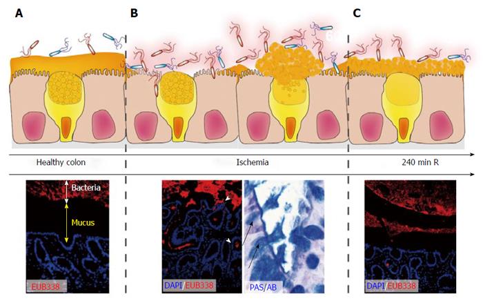
Apart from the epithelial lining, a crucial layer of antimicrobial defense, particularly in the colon, is the mucus layer. This layer creates a physical and chemical barrier that prevents adherence of microbiota to the epithelium (Figure 4A)[19-21]. To study the consequences of IR on the mucus layer in more detail, implementation of a rat colon IR model was imperative to overcome problems with preservation of the mucus layer and staining for bacteria.

Figure 4
Figure 4 Rapid restoration of colon ischemia-reperfusion-induced mucus barrier loss by goblet cell compound exocytosis. In healthy colon, bacteria are separated from the epithelium by a thick mucus layer (A; DAPI, Blue: nuclei. EUB338, Red: bacteria); During ischemia and early reperfusion, the mucus barrier is damaged leading to penetration of bacteria deep into the colonic crypts (B). Goblet cells respond by massive release of their granules into the crypt lumen (so called compound exocytosis), which led to a recoverd mucus layer at 240 min of reperfusion in the rat intestine (C). Note that bacterial clearance is accompanied by depletion of goblet cell contents.

A newly observed and likely important pathophysiological consequence of 60 min of colonic ischemia was disruption of this mucus layer, which was accompanied by intrusion of bacteria into the colonic epithelium (Figure 4B). Intriguingly, goblet cells react rapidly in response to these bacterial threats with expulsion of their mucus - containing granules (Figure 4B, right panel), a phenomenon known as goblet cell “compound exocytosis”[21,75]. Compound exocytosis is characterized by expansion of mucin volume and rapid replacement of degraded mucus barriers, and has been described as a mechanism to reinforce the barrier and exclude pathogens from the normally sterile crypts[21]. Indeed, the mucus layer covering the epithelium was fully restored within 4 h of reperfusion of ischemically damaged colon (Figure 4C) and a time-dependent clearance of bacteria from the colonic crypts was observed. This was accompanied by only mild inflammatory responses, particularly if compared to small intestinal IR[73].

Therefore, compound exocytosis seemed an efficient mechanism to prevent prolonged exposure of microbiota to the epithelium thereby preventing intestinal inflammation. This might explain why patients with colonic ischemia tend to have a milder course of the disease as compared to small intestinal IR[71,76-78]. In addition, mucus organization could explain why ischemia of the right-sided colon is often more severe than left-sided colonic ischemia, since thickness of the inner mucus layer increases from the proximal to distal colon[71].

TRANSLATION TO CLINICAL PRACTICE

Our improved understanding of the pathophysiological mechanisms of small intestinal IR paves a way to develop and experimentally test new therapies that can limit ischemic damage as well as reperfusion-induced intestinal tissue damage. This is particularly relevant for patients undergoing aortic surgery with temporary abrogation of the mesenteric blood flow, patients with severe mesenteric hypoperfusion due to sepsis or shock, or patients revascularized after an ischemic insult (i.e., after embolectomy). In addition, improved knowledge on the sequelae of human intestinal IR is important for the field of intestinal transplantation.

However, probably the most important factor that determines the outcome of patients with acute intestinal ischemia is our ability to recognize intestinal ischemia at an early stage[14]. There is often a considerable diagnostic delay due to the nonspecific clinical presentation in combination with the lack of early, non-invasive diagnostic markers for intestinal ischemia. A promising marker for intestinal ischemia is intestinal fatty acid binding protein (I-FABP), a small (14-kD) cytosolic protein specifically present in mature enterocytes at the tip of the villus that is released into the circulation upon enterocyte damage[5,79-82]. Using the human intestinal IR model, the characteristics of I-FABP were studied in more detail. We have shown that I-FABP is a very sensitive marker of intestinal villus tip damage, as arteriovenous concentration differences were already increased 15 min after onset of ischemia, whereas morphological changes were hardly noticeable at this time point[83]. Longer ischemia resulted in massive increase of arteriovenous I-FABP concentration differences, and I-FABP levels correlated with the degree of histological epithelial damage.

More importantly however, systemic I-FABP levels were increased in patients that were included in the experimental IR protocol compared to control patients that were exposed to similar surgical handling without being exposed to intestinal IR using the experimental protocol[83]. This demonstrated that IR of a 6 centimeter small intestinal segment can be detected using plasma I-FABP measurement, which emphasizes the potential value of plasma I-FABP in the early diagnosis of intestinal ischemia. Prospective trials now need to show whether I-FABP levels can be used for decision making in patients with suspected intestinal ischemia.

CONCLUSION

Over the past years, the human experimental intestinal IR models have led to a more in depth knowledge on the pathophysiology of human small intestinal IR and colon IR. We have been able to identify the duration of ischemia that the human small intestine can undergo before extensive physical and immunological barrier damage and inflammation occur. Our studies show that the human small intestine is adapted to withstand short periods of ischemia by ingenious mechanisms involved in prevention and rapid repair of epithelial lining damage, thereby preventing severe inflammation. This explains why humans can endure short periods of intestinal IR as a consequence of strenuous exercise (physiological), or during major (cardiovascular) surgery with cross clamping of the aorta or extensive blood loss. In contrast, longer periods of ischemia result in extended physical barrier damage and immunological barrier compromise, typically observed in the small intestine.

The colonic epithelial lining appeared more resistant to ischemia and reperfusion, and goblet cells reacted rapidly to IR-induced mucus barrier loss by expulsion of their contents, which resulted in rapid restoration of the protective mucus barrier.

With increasing knowledge on the pathophysiology of human intestinal IR, future studies will be directed towards implementation of this knowledge on intestinal IR, to work towards better detection, prevention and therapy for intestinal ischemia/reperfusion in man.

Footnotes

P- Reviewer: Karhausen J, Zhao L S- Editor: Yu J L- Editor: A E- Editor: Wang CH

References
1.  American Gastroenterological Association Medical Position Statement: guidelines on intestinal ischemia Gastroenterology. 2000;118:951-953.  [PubMed]  [DOI]  [Cited in This Article: ]  [Cited by in Crossref: 105]  [Cited by in F6Publishing: 80]  [Article Influence: 3.3]  [Reference Citation Analysis (0)]
2.  Bradbury AW, Brittenden J, McBride K, Ruckley CV. Mesenteric ischaemia: a multidisciplinary approach. Br J Surg. 1995;82:1446-1459.  [PubMed]  [DOI]  [Cited in This Article: ]  [Cited by in Crossref: 171]  [Cited by in F6Publishing: 177]  [Article Influence: 6.1]  [Reference Citation Analysis (0)]
3.  Perko MJ, Nielsen HB, Skak C, Clemmesen JO, Schroeder TV, Secher NH. Mesenteric, coeliac and splanchnic blood flow in humans during exercise. J Physiol. 1998;513:907-913.  [PubMed]  [DOI]  [Cited in This Article: ]  [Cited by in Crossref: 98]  [Cited by in F6Publishing: 98]  [Article Influence: 3.8]  [Reference Citation Analysis (0)]
4.  van Wijck K, Lenaerts K, van Loon LJ, Peters WH, Buurman WA, Dejong CH. Exercise-induced splanchnic hypoperfusion results in gut dysfunction in healthy men. PLoS One. 2011;6:e22366.  [PubMed]  [DOI]  [Cited in This Article: ]  [Cited by in Crossref: 193]  [Cited by in F6Publishing: 226]  [Article Influence: 17.4]  [Reference Citation Analysis (0)]
5.  Derikx JP, Matthijsen RA, de Bruïne AP, van Bijnen AA, Heineman E, van Dam RM, Dejong CH, Buurman WA. Rapid reversal of human intestinal ischemia-reperfusion induced damage by shedding of injured enterocytes and reepithelialisation. PLoS One. 2008;3:e3428.  [PubMed]  [DOI]  [Cited in This Article: ]  [Cited by in Crossref: 73]  [Cited by in F6Publishing: 82]  [Article Influence: 5.1]  [Reference Citation Analysis (0)]
6.  Blikslager AT, Moeser AJ, Gookin JL, Jones SL, Odle J. Restoration of barrier function in injured intestinal mucosa. Physiol Rev. 2007;87:545-564.  [PubMed]  [DOI]  [Cited in This Article: ]  [Cited by in Crossref: 375]  [Cited by in F6Publishing: 409]  [Article Influence: 24.1]  [Reference Citation Analysis (0)]
7.  Colgan SP, Taylor CT. Hypoxia: an alarm signal during intestinal inflammation. Nat Rev Gastroenterol Hepatol. 2010;7:281-287.  [PubMed]  [DOI]  [Cited in This Article: ]  [Cited by in Crossref: 313]  [Cited by in F6Publishing: 354]  [Article Influence: 25.3]  [Reference Citation Analysis (0)]
8.  Hatoum OA, Binion DG, Otterson MF, Gutterman DD. Acquired microvascular dysfunction in inflammatory bowel disease: Loss of nitric oxide-mediated vasodilation. Gastroenterology. 2003;125:58-69.  [PubMed]  [DOI]  [Cited in This Article: ]  [Cited by in Crossref: 185]  [Cited by in F6Publishing: 193]  [Article Influence: 9.2]  [Reference Citation Analysis (0)]
9.  Mensink PB, Moons LM, Kuipers EJ. Chronic gastrointestinal ischaemia: shifting paradigms. Gut. 2011;60:722-737.  [PubMed]  [DOI]  [Cited in This Article: ]  [Cited by in Crossref: 61]  [Cited by in F6Publishing: 62]  [Article Influence: 4.8]  [Reference Citation Analysis (0)]
10.  Derrick JR, Pollard HS, Moore RM. The pattern of arteriosclerotic narrowing of the celiac and superior mesenteric arteries. Ann Surg. 1959;149:684-689.  [PubMed]  [DOI]  [Cited in This Article: ]  [Cited by in Crossref: 109]  [Cited by in F6Publishing: 102]  [Article Influence: 4.3]  [Reference Citation Analysis (0)]
11.  Oldenburg WA, Lau LL, Rodenberg TJ, Edmonds HJ, Burger CD. Acute mesenteric ischemia: a clinical review. Arch Intern Med. 2004;164:1054-1062.  [PubMed]  [DOI]  [Cited in This Article: ]  [Cited by in Crossref: 444]  [Cited by in F6Publishing: 415]  [Article Influence: 20.8]  [Reference Citation Analysis (0)]
12.  Kozuch PL, Brandt LJ. Review article: diagnosis and management of mesenteric ischaemia with an emphasis on pharmacotherapy. Aliment Pharmacol Ther. 2005;21:201-215.  [PubMed]  [DOI]  [Cited in This Article: ]  [Cited by in Crossref: 118]  [Cited by in F6Publishing: 121]  [Article Influence: 6.4]  [Reference Citation Analysis (0)]
13.  Kassahun WT, Schulz T, Richter O, Hauss J. Unchanged high mortality rates from acute occlusive intestinal ischemia: six year review. Langenbecks Arch Surg. 2008;393:163-171.  [PubMed]  [DOI]  [Cited in This Article: ]  [Cited by in Crossref: 171]  [Cited by in F6Publishing: 159]  [Article Influence: 9.9]  [Reference Citation Analysis (0)]
14.  Kougias P, Lau D, El Sayed HF, Zhou W, Huynh TT, Lin PH. Determinants of mortality and treatment outcome following surgical interventions for acute mesenteric ischemia. J Vasc Surg. 2007;46:467-474.  [PubMed]  [DOI]  [Cited in This Article: ]  [Cited by in Crossref: 192]  [Cited by in F6Publishing: 152]  [Article Influence: 8.9]  [Reference Citation Analysis (0)]
15.  Homer-Vanniasinkam S, Crinnion JN, Gough MJ. Post-ischaemic organ dysfunction: a review. Eur J Vasc Endovasc Surg. 1997;14:195-203.  [PubMed]  [DOI]  [Cited in This Article: ]  [Cited by in Crossref: 79]  [Cited by in F6Publishing: 85]  [Article Influence: 3.1]  [Reference Citation Analysis (0)]
16.  Schoenberg MH, Beger HG. Reperfusion injury after intestinal ischemia. Crit Care Med. 1993;21:1376-1386.  [PubMed]  [DOI]  [Cited in This Article: ]  [Cited by in Crossref: 214]  [Cited by in F6Publishing: 199]  [Article Influence: 6.4]  [Reference Citation Analysis (0)]
17.  Sansonetti PJ. War and peace at mucosal surfaces. Nat Rev Immunol. 2004;4:953-964.  [PubMed]  [DOI]  [Cited in This Article: ]  [Cited by in Crossref: 474]  [Cited by in F6Publishing: 465]  [Article Influence: 24.5]  [Reference Citation Analysis (0)]
18.  Grootjans J, Thuijls G, Verdam F, Derikx JP, Lenaerts K, Buurman WA. Non-invasive assessment of barrier integrity and function of the human gut. World J Gastrointest Surg. 2010;2:61-69.  [PubMed]  [DOI]  [Cited in This Article: ]  [Cited by in CrossRef: 137]  [Cited by in F6Publishing: 146]  [Article Influence: 10.4]  [Reference Citation Analysis (0)]
19.  Deplancke B, Gaskins HR. Microbial modulation of innate defense: goblet cells and the intestinal mucus layer. Am J Clin Nutr. 2001;73:1131S-1141S.  [PubMed]  [DOI]  [Cited in This Article: ]
20.  Johansson ME, Phillipson M, Petersson J, Velcich A, Holm L, Hansson GC. The inner of the two Muc2 mucin-dependent mucus layers in colon is devoid of bacteria. Proc Natl Acad Sci USA. 2008;105:15064-15069.  [PubMed]  [DOI]  [Cited in This Article: ]  [Cited by in Crossref: 1326]  [Cited by in F6Publishing: 1454]  [Article Influence: 90.9]  [Reference Citation Analysis (1)]
21.  McGuckin MA, Lindén SK, Sutton P, Florin TH. Mucin dynamics and enteric pathogens. Nat Rev Microbiol. 2011;9:265-278.  [PubMed]  [DOI]  [Cited in This Article: ]  [Cited by in Crossref: 907]  [Cited by in F6Publishing: 966]  [Article Influence: 74.3]  [Reference Citation Analysis (0)]
22.  Bjarnason I, MacPherson A, Hollander D. Intestinal permeability: an overview. Gastroenterology. 1995;108:1566-1581.  [PubMed]  [DOI]  [Cited in This Article: ]  [Cited by in Crossref: 627]  [Cited by in F6Publishing: 623]  [Article Influence: 21.5]  [Reference Citation Analysis (0)]
23.  Koch S, Nusrat A. The life and death of epithelia during inflammation: lessons learned from the gut. Annu Rev Pathol. 2012;7:35-60.  [PubMed]  [DOI]  [Cited in This Article: ]  [Cited by in Crossref: 109]  [Cited by in F6Publishing: 101]  [Article Influence: 7.8]  [Reference Citation Analysis (0)]
24.  Turner JR. Intestinal mucosal barrier function in health and disease. Nat Rev Immunol. 2009;9:799-809.  [PubMed]  [DOI]  [Cited in This Article: ]  [Cited by in Crossref: 2121]  [Cited by in F6Publishing: 2541]  [Article Influence: 169.4]  [Reference Citation Analysis (0)]
25.  Nagler-Anderson C. Man the barrier! Strategic defences in the intestinal mucosa. Nat Rev Immunol. 2001;1:59-67.  [PubMed]  [DOI]  [Cited in This Article: ]  [Cited by in Crossref: 243]  [Cited by in F6Publishing: 205]  [Article Influence: 8.9]  [Reference Citation Analysis (0)]
26.  Medzhitov R, Janeway CA. Decoding the patterns of self and nonself by the innate immune system. Science. 2002;296:298-300.  [PubMed]  [DOI]  [Cited in This Article: ]  [Cited by in Crossref: 1534]  [Cited by in F6Publishing: 1422]  [Article Influence: 64.6]  [Reference Citation Analysis (0)]
27.  Selsted ME, Ouellette AJ. Mammalian defensins in the antimicrobial immune response. Nat Immunol. 2005;6:551-557.  [PubMed]  [DOI]  [Cited in This Article: ]  [Cited by in Crossref: 874]  [Cited by in F6Publishing: 859]  [Article Influence: 45.2]  [Reference Citation Analysis (0)]
28.  Salzman NH, Hung K, Haribhai D, Chu H, Karlsson-Sjöberg J, Amir E, Teggatz P, Barman M, Hayward M, Eastwood D. Enteric defensins are essential regulators of intestinal microbial ecology. Nat Immunol. 2010;11:76-83.  [PubMed]  [DOI]  [Cited in This Article: ]  [Cited by in Crossref: 835]  [Cited by in F6Publishing: 880]  [Article Influence: 58.7]  [Reference Citation Analysis (0)]
29.  Ouellette AJ. Paneth cells and innate mucosal immunity. Curr Opin Gastroenterol. 2010;26:547-553.  [PubMed]  [DOI]  [Cited in This Article: ]  [Cited by in Crossref: 118]  [Cited by in F6Publishing: 109]  [Article Influence: 7.8]  [Reference Citation Analysis (0)]
30.  Bevins CL, Salzman NH. Paneth cells, antimicrobial peptides and maintenance of intestinal homeostasis. Nat Rev Microbiol. 2011;9:356-368.  [PubMed]  [DOI]  [Cited in This Article: ]  [Cited by in Crossref: 725]  [Cited by in F6Publishing: 829]  [Article Influence: 63.8]  [Reference Citation Analysis (0)]
31.  Ayabe T, Satchell DP, Wilson CL, Parks WC, Selsted ME, Ouellette AJ. Secretion of microbicidal alpha-defensins by intestinal Paneth cells in response to bacteria. Nat Immunol. 2000;1:113-118.  [PubMed]  [DOI]  [Cited in This Article: ]  [Cited by in Crossref: 776]  [Cited by in F6Publishing: 757]  [Article Influence: 31.5]  [Reference Citation Analysis (0)]
32.  Mukherjee S, Vaishnava S, Hooper LV. Multi-layered regulation of intestinal antimicrobial defense. Cell Mol Life Sci. 2008;65:3019-3027.  [PubMed]  [DOI]  [Cited in This Article: ]  [Cited by in Crossref: 107]  [Cited by in F6Publishing: 101]  [Article Influence: 6.3]  [Reference Citation Analysis (0)]
33.  Vaishnava S, Behrendt CL, Ismail AS, Eckmann L, Hooper LV. Paneth cells directly sense gut commensals and maintain homeostasis at the intestinal host-microbial interface. Proc Natl Acad Sci USA. 2008;105:20858-20863.  [PubMed]  [DOI]  [Cited in This Article: ]  [Cited by in Crossref: 715]  [Cited by in F6Publishing: 750]  [Article Influence: 46.9]  [Reference Citation Analysis (0)]
34.  Grootjans J, Hodin CM, de Haan JJ, Derikx JP, Rouschop KM, Verheyen FK, van Dam RM, Dejong CH, Buurman WA, Lenaerts K. Level of activation of the unfolded protein response correlates with Paneth cell apoptosis in human small intestine exposed to ischemia/reperfusion. Gastroenterology. 2011;140:529-539.e3.  [PubMed]  [DOI]  [Cited in This Article: ]
35.  Meyer-Hoffert U, Hornef MW, Henriques-Normark B, Axelsson LG, Midtvedt T, Pütsep K, Andersson M. Secreted enteric antimicrobial activity localises to the mucus surface layer. Gut. 2008;57:764-771.  [PubMed]  [DOI]  [Cited in This Article: ]  [Cited by in Crossref: 209]  [Cited by in F6Publishing: 208]  [Article Influence: 13.0]  [Reference Citation Analysis (0)]
36.  Sato T, van Es JH, Snippert HJ, Stange DE, Vries RG, van den Born M, Barker N, Shroyer NF, van de Wetering M, Clevers H. Paneth cells constitute the niche for Lgr5 stem cells in intestinal crypts. Nature. 2011;469:415-418.  [PubMed]  [DOI]  [Cited in This Article: ]  [Cited by in Crossref: 1728]  [Cited by in F6Publishing: 1844]  [Article Influence: 131.7]  [Reference Citation Analysis (0)]
37.  Gonzalez LM, Moeser AJ, Blikslager AT. Animal models of ischemia-reperfusion-induced intestinal injury: progress and promise for translational research. Am J Physiol Gastrointest Liver Physiol. 2015;308:G63-G75.  [PubMed]  [DOI]  [Cited in This Article: ]  [Cited by in Crossref: 132]  [Cited by in F6Publishing: 172]  [Article Influence: 19.1]  [Reference Citation Analysis (0)]
38.  Derikx JP, Matthijsen RA, de Bruïne AP, van Dam RM, Buurman WA, Dejong CH. A new model to study intestinal ischemia-reperfusion damage in man. J Surg Res. 2011;166:222-226.  [PubMed]  [DOI]  [Cited in This Article: ]  [Cited by in Crossref: 20]  [Cited by in F6Publishing: 20]  [Article Influence: 1.3]  [Reference Citation Analysis (0)]
39.  Grootjans J, Thuijls G, Derikx JP, van Dam RM, Dejong CH, Buurman WA. Rapid lamina propria retraction and zipper-like constriction of the epithelium preserves the epithelial lining in human small intestine exposed to ischaemia-reperfusion. J Pathol. 2011;224:411-419.  [PubMed]  [DOI]  [Cited in This Article: ]  [Cited by in Crossref: 22]  [Cited by in F6Publishing: 24]  [Article Influence: 1.8]  [Reference Citation Analysis (0)]
40.  Chiu CJ, McArdle AH, Brown R, Scott HJ, Gurd FN. Intestinal mucosal lesion in low-flow states. I. A morphological, hemodynamic, and metabolic reappraisal. Arch Surg. 1970;101:478-483.  [PubMed]  [DOI]  [Cited in This Article: ]  [Cited by in Crossref: 1258]  [Cited by in F6Publishing: 1364]  [Article Influence: 25.3]  [Reference Citation Analysis (0)]
41.  Park PO, Haglund U, Bulkley GB, Fält K. The sequence of development of intestinal tissue injury after strangulation ischemia and reperfusion. Surgery. 1990;107:574-580.  [PubMed]  [DOI]  [Cited in This Article: ]
42.  Gassler N, Roth W, Funke B, Schneider A, Herzog F, Tischendorf JJ, Grund K, Penzel R, Bravo IG, Mariadason J. Regulation of enterocyte apoptosis by acyl-CoA synthetase 5 splicing. Gastroenterology. 2007;133:587-598.  [PubMed]  [DOI]  [Cited in This Article: ]  [Cited by in Crossref: 38]  [Cited by in F6Publishing: 43]  [Article Influence: 2.5]  [Reference Citation Analysis (0)]
43.  Bullen TF, Forrest S, Campbell F, Dodson AR, Hershman MJ, Pritchard DM, Turner JR, Montrose MH, Watson AJ. Characterization of epithelial cell shedding from human small intestine. Lab Invest. 2006;86:1052-1063.  [PubMed]  [DOI]  [Cited in This Article: ]  [Cited by in Crossref: 155]  [Cited by in F6Publishing: 141]  [Article Influence: 7.8]  [Reference Citation Analysis (0)]
44.  Russo JM, Florian P, Shen L, Graham WV, Tretiakova MS, Gitter AH, Mrsny RJ, Turner JR. Distinct temporal-spatial roles for rho kinase and myosin light chain kinase in epithelial purse-string wound closure. Gastroenterology. 2005;128:987-1001.  [PubMed]  [DOI]  [Cited in This Article: ]  [Cited by in Crossref: 101]  [Cited by in F6Publishing: 104]  [Article Influence: 5.5]  [Reference Citation Analysis (0)]
45.  Martin P, Parkhurst SM. Parallels between tissue repair and embryo morphogenesis. Development. 2004;131:3021-3034.  [PubMed]  [DOI]  [Cited in This Article: ]  [Cited by in Crossref: 417]  [Cited by in F6Publishing: 400]  [Article Influence: 20.0]  [Reference Citation Analysis (0)]
46.  Nusrat A, Delp C, Madara JL. Intestinal epithelial restitution. Characterization of a cell culture model and mapping of cytoskeletal elements in migrating cells. J Clin Invest. 1992;89:1501-1511.  [PubMed]  [DOI]  [Cited in This Article: ]  [Cited by in Crossref: 181]  [Cited by in F6Publishing: 178]  [Article Influence: 5.6]  [Reference Citation Analysis (0)]
47.  Florian P, Schöneberg T, Schulzke JD, Fromm M, Gitter AH. Single-cell epithelial defects close rapidly by an actinomyosin purse string mechanism with functional tight junctions. J Physiol. 2002;545:485-499.  [PubMed]  [DOI]  [Cited in This Article: ]  [Cited by in Crossref: 57]  [Cited by in F6Publishing: 60]  [Article Influence: 2.7]  [Reference Citation Analysis (0)]
48.  Matthijsen RA, Derikx JP, Kuipers D, van Dam RM, Dejong CH, Buurman WA. Enterocyte shedding and epithelial lining repair following ischemia of the human small intestine attenuate inflammation. PLoS One. 2009;4:e7045.  [PubMed]  [DOI]  [Cited in This Article: ]  [Cited by in Crossref: 43]  [Cited by in F6Publishing: 47]  [Article Influence: 3.1]  [Reference Citation Analysis (0)]
49.  Grootjans J, Lenaerts K, Derikx JP, Matthijsen RA, de Bruïne AP, van Bijnen AA, van Dam RM, Dejong CH, Buurman WA. Human intestinal ischemia-reperfusion-induced inflammation characterized: experiences from a new translational model. Am J Pathol. 2010;176:2283-2291.  [PubMed]  [DOI]  [Cited in This Article: ]  [Cited by in Crossref: 110]  [Cited by in F6Publishing: 126]  [Article Influence: 9.0]  [Reference Citation Analysis (0)]
50.  Blikslager AT, Roberts MC, Rhoads JM, Argenzio RA. Is reperfusion injury an important cause of mucosal damage after porcine intestinal ischemia? Surgery. 1997;121:526-534.  [PubMed]  [DOI]  [Cited in This Article: ]  [Cited by in Crossref: 47]  [Cited by in F6Publishing: 53]  [Article Influence: 2.0]  [Reference Citation Analysis (0)]
51.  Boxer L, Dale DC. Neutropenia: causes and consequences. Semin Hematol. 2002;39:75-81.  [PubMed]  [DOI]  [Cited in This Article: ]  [Cited by in Crossref: 80]  [Cited by in F6Publishing: 75]  [Article Influence: 3.4]  [Reference Citation Analysis (0)]
52.  Matthijsen RA, Huugen D, Hoebers NT, de Vries B, Peutz-Kootstra CJ, Aratani Y, Daha MR, Tervaert JW, Buurman WA, Heeringa P. Myeloperoxidase is critically involved in the induction of organ damage after renal ischemia reperfusion. Am J Pathol. 2007;171:1743-1752.  [PubMed]  [DOI]  [Cited in This Article: ]  [Cited by in Crossref: 90]  [Cited by in F6Publishing: 92]  [Article Influence: 5.4]  [Reference Citation Analysis (0)]
53.  Schoenberg MH, Poch B, Younes M, Schwarz A, Baczako K, Lundberg C, Haglund U, Beger HG. Involvement of neutrophils in postischaemic damage to the small intestine. Gut. 1991;32:905-912.  [PubMed]  [DOI]  [Cited in This Article: ]  [Cited by in Crossref: 87]  [Cited by in F6Publishing: 79]  [Article Influence: 2.4]  [Reference Citation Analysis (0)]
54.  Arumugam TV, Shiels IA, Woodruff TM, Granger DN, Taylor SM. The role of the complement system in ischemia-reperfusion injury. Shock. 2004;21:401-409.  [PubMed]  [DOI]  [Cited in This Article: ]  [Cited by in Crossref: 229]  [Cited by in F6Publishing: 233]  [Article Influence: 11.7]  [Reference Citation Analysis (0)]
55.  Woodcock SA, Kyriakides C, Wang Y, Austen WG, Moore FD, Valeri R, Hartwell D, Hechtman HB. Soluble P-selectin moderates complement dependent injury. Shock. 2000;14:610-615.  [PubMed]  [DOI]  [Cited in This Article: ]  [Cited by in Crossref: 10]  [Cited by in F6Publishing: 10]  [Article Influence: 0.4]  [Reference Citation Analysis (0)]
56.  Zhang M, Austen WG, Chiu I, Alicot EM, Hung R, Ma M, Verna N, Xu M, Hechtman HB, Moore FD. Identification of a specific self-reactive IgM antibody that initiates intestinal ischemia/reperfusion injury. Proc Natl Acad Sci USA. 2004;101:3886-3891.  [PubMed]  [DOI]  [Cited in This Article: ]  [Cited by in Crossref: 178]  [Cited by in F6Publishing: 182]  [Article Influence: 9.1]  [Reference Citation Analysis (0)]
57.  Stahl GL, Xu Y, Hao L, Miller M, Buras JA, Fung M, Zhao H. Role for the alternative complement pathway in ischemia/reperfusion injury. Am J Pathol. 2003;162:449-455.  [PubMed]  [DOI]  [Cited in This Article: ]  [Cited by in Crossref: 127]  [Cited by in F6Publishing: 130]  [Article Influence: 6.2]  [Reference Citation Analysis (0)]
58.  Hart ML, Ceonzo KA, Shaffer LA, Takahashi K, Rother RP, Reenstra WR, Buras JA, Stahl GL. Gastrointestinal ischemia-reperfusion injury is lectin complement pathway dependent without involving C1q. J Immunol. 2005;174:6373-6380.  [PubMed]  [DOI]  [Cited in This Article: ]  [Cited by in Crossref: 156]  [Cited by in F6Publishing: 158]  [Article Influence: 8.3]  [Reference Citation Analysis (0)]
59.  Thurman JM, Lenderink AM, Royer PA, Coleman KE, Zhou J, Lambris JD, Nemenoff RA, Quigg RJ, Holers VM. C3a is required for the production of CXC chemokines by tubular epithelial cells after renal ishemia/reperfusion. J Immunol. 2007;178:1819-1828.  [PubMed]  [DOI]  [Cited in This Article: ]  [Cited by in Crossref: 82]  [Cited by in F6Publishing: 89]  [Article Influence: 5.2]  [Reference Citation Analysis (0)]
60.  Kaser A, Lee AH, Franke A, Glickman JN, Zeissig S, Tilg H, Nieuwenhuis EE, Higgins DE, Schreiber S, Glimcher LH. XBP1 links ER stress to intestinal inflammation and confers genetic risk for human inflammatory bowel disease. Cell. 2008;134:743-756.  [PubMed]  [DOI]  [Cited in This Article: ]  [Cited by in Crossref: 1014]  [Cited by in F6Publishing: 1101]  [Article Influence: 68.8]  [Reference Citation Analysis (0)]
61.  Kaser A, Blumberg RS. Endoplasmic reticulum stress and intestinal inflammation. Mucosal Immunol. 2010;3:11-16.  [PubMed]  [DOI]  [Cited in This Article: ]  [Cited by in Crossref: 99]  [Cited by in F6Publishing: 105]  [Article Influence: 7.5]  [Reference Citation Analysis (0)]
62.  Koritzinsky M, Magagnin MG, van den Beucken T, Seigneuric R, Savelkouls K, Dostie J, Pyronnet S, Kaufman RJ, Weppler SA, Voncken JW. Gene expression during acute and prolonged hypoxia is regulated by distinct mechanisms of translational control. EMBO J. 2006;25:1114-1125.  [PubMed]  [DOI]  [Cited in This Article: ]  [Cited by in Crossref: 268]  [Cited by in F6Publishing: 273]  [Article Influence: 15.2]  [Reference Citation Analysis (0)]
63.  Xu C, Bailly-Maitre B, Reed JC. Endoplasmic reticulum stress: cell life and death decisions. J Clin Invest. 2005;115:2656-2664.  [PubMed]  [DOI]  [Cited in This Article: ]  [Cited by in Crossref: 1683]  [Cited by in F6Publishing: 1815]  [Article Influence: 95.5]  [Reference Citation Analysis (0)]
64.  Szegezdi E, Logue SE, Gorman AM, Samali A. Mediators of endoplasmic reticulum stress-induced apoptosis. EMBO Rep. 2006;7:880-885.  [PubMed]  [DOI]  [Cited in This Article: ]  [Cited by in Crossref: 1670]  [Cited by in F6Publishing: 1843]  [Article Influence: 102.4]  [Reference Citation Analysis (0)]
65.  Montie HL, Kayali F, Haezebrouck AJ, Rossi NF, Degracia DJ. Renal ischemia and reperfusion activates the eIF 2 alpha kinase PERK. Biochim Biophys Acta. 2005;1741:314-324.  [PubMed]  [DOI]  [Cited in This Article: ]  [Cited by in Crossref: 31]  [Cited by in F6Publishing: 36]  [Article Influence: 2.0]  [Reference Citation Analysis (0)]
66.  Yung HW, Korolchuk S, Tolkovsky AM, Charnock-Jones DS, Burton GJ. Endoplasmic reticulum stress exacerbates ischemia-reperfusion-induced apoptosis through attenuation of Akt protein synthesis in human choriocarcinoma cells. FASEB J. 2007;21:872-884.  [PubMed]  [DOI]  [Cited in This Article: ]  [Cited by in Crossref: 99]  [Cited by in F6Publishing: 102]  [Article Influence: 5.7]  [Reference Citation Analysis (0)]
67.  DeGracia DJ. Acute and persistent protein synthesis inhibition following cerebral reperfusion. J Neurosci Res. 2004;77:771-776.  [PubMed]  [DOI]  [Cited in This Article: ]  [Cited by in Crossref: 33]  [Cited by in F6Publishing: 33]  [Article Influence: 1.7]  [Reference Citation Analysis (0)]
68.  Adolph TE, Tomczak MF, Niederreiter L, Ko HJ, Böck J, Martinez-Naves E, Glickman JN, Tschurtschenthaler M, Hartwig J, Hosomi S. Paneth cells as a site of origin for intestinal inflammation. Nature. 2013;503:272-276.  [PubMed]  [DOI]  [Cited in This Article: ]  [Cited by in Crossref: 584]  [Cited by in F6Publishing: 552]  [Article Influence: 50.2]  [Reference Citation Analysis (0)]
69.  Longstreth GF, Yao JF. Epidemiology, clinical features, high-risk factors, and outcome of acute large bowel ischemia. Clin Gastroenterol Hepatol. 2009;7:1075-1080.e1-2; quiz 1023.  [PubMed]  [DOI]  [Cited in This Article: ]
70.  Feuerstadt P, Brandt LJ. Colon ischemia: recent insights and advances. Curr Gastroenterol Rep. 2010;12:383-390.  [PubMed]  [DOI]  [Cited in This Article: ]  [Cited by in Crossref: 49]  [Cited by in F6Publishing: 54]  [Article Influence: 3.9]  [Reference Citation Analysis (0)]
71.  Brandt LJ, Feuerstadt P, Blaszka MC. Anatomic patterns, patient characteristics, and clinical outcomes in ischemic colitis: a study of 313 cases supported by histology. Am J Gastroenterol. 2010;105:2245-2252; quiz 2253.  [PubMed]  [DOI]  [Cited in This Article: ]  [Cited by in Crossref: 136]  [Cited by in F6Publishing: 133]  [Article Influence: 9.5]  [Reference Citation Analysis (0)]
72.  Grootjans J, Hundscheid IH, Buurman WA. Goblet cell compound exocytosis in the defense against bacterial invasion in the colon exposed to ischemia-reperfusion. Gut Microbes. 2013;4:232-235.  [PubMed]  [DOI]  [Cited in This Article: ]  [Cited by in Crossref: 11]  [Cited by in F6Publishing: 16]  [Article Influence: 1.5]  [Reference Citation Analysis (0)]
73.  Grootjans J, Hundscheid IH, Lenaerts K, Boonen B, Renes IB, Verheyen FK, Dejong CH, von Meyenfeldt MF, Beets GL, Buurman WA. Ischaemia-induced mucus barrier loss and bacterial penetration are rapidly counteracted by increased goblet cell secretory activity in human and rat colon. Gut. 2013;62:250-258.  [PubMed]  [DOI]  [Cited in This Article: ]  [Cited by in Crossref: 70]  [Cited by in F6Publishing: 77]  [Article Influence: 7.0]  [Reference Citation Analysis (0)]
74.  Hundscheid IH, Grootjans J, Lenaerts K, Schellekens DH, Derikx JP, Boonen BT, von Meyenfeldt MF, Beets GL, Buurman WA, Dejong CH. The Human Colon Is More Resistant to Ischemia-reperfusion-induced Tissue Damage Than the Small Intestine: An Observational Study. Ann Surg. 2015;262:304-311.  [PubMed]  [DOI]  [Cited in This Article: ]  [Cited by in Crossref: 11]  [Cited by in F6Publishing: 13]  [Article Influence: 1.4]  [Reference Citation Analysis (0)]
75.  Davis CW, Dickey BF. Regulated airway goblet cell mucin secretion. Annu Rev Physiol. 2008;70:487-512.  [PubMed]  [DOI]  [Cited in This Article: ]  [Cited by in Crossref: 176]  [Cited by in F6Publishing: 186]  [Article Influence: 11.6]  [Reference Citation Analysis (0)]
76.  Montoro MA, Brandt LJ, Santolaria S, Gomollon F, Sánchez Puértolas B, Vera J, Bujanda L, Cosme A, Cabriada JL, Durán M. Clinical patterns and outcomes of ischaemic colitis: results of the Working Group for the Study of Ischaemic Colitis in Spain (CIE study). Scand J Gastroenterol. 2011;46:236-246.  [PubMed]  [DOI]  [Cited in This Article: ]  [Cited by in Crossref: 101]  [Cited by in F6Publishing: 107]  [Article Influence: 8.2]  [Reference Citation Analysis (0)]
77.  Elder K, Lashner BA, Al Solaiman F. Clinical approach to colonic ischemia. Cleve Clin J Med. 2009;76:401-409.  [PubMed]  [DOI]  [Cited in This Article: ]  [Cited by in Crossref: 65]  [Cited by in F6Publishing: 68]  [Article Influence: 4.5]  [Reference Citation Analysis (1)]
78.  Green BT, Tendler DA. Ischemic colitis: a clinical review. South Med J. 2005;98:217-222.  [PubMed]  [DOI]  [Cited in This Article: ]  [Cited by in Crossref: 117]  [Cited by in F6Publishing: 105]  [Article Influence: 5.5]  [Reference Citation Analysis (0)]
79.  Pelsers MM, Hermens WT, Glatz JF. Fatty acid-binding proteins as plasma markers of tissue injury. Clin Chim Acta. 2005;352:15-35.  [PubMed]  [DOI]  [Cited in This Article: ]  [Cited by in Crossref: 317]  [Cited by in F6Publishing: 332]  [Article Influence: 17.5]  [Reference Citation Analysis (0)]
80.  Derikx JP, Vreugdenhil AC, Van den Neucker AM, Grootjans J, van Bijnen AA, Damoiseaux JG, van Heurn LW, Heineman E, Buurman WA. A pilot study on the noninvasive evaluation of intestinal damage in celiac disease using I-FABP and L-FABP. J Clin Gastroenterol. 2009;43:727-733.  [PubMed]  [DOI]  [Cited in This Article: ]  [Cited by in Crossref: 69]  [Cited by in F6Publishing: 77]  [Article Influence: 5.1]  [Reference Citation Analysis (0)]
81.  Lieberman JM, Sacchettini J, Marks C, Marks WH. Human intestinal fatty acid binding protein: report of an assay with studies in normal volunteers and intestinal ischemia. Surgery. 1997;121:335-342.  [PubMed]  [DOI]  [Cited in This Article: ]  [Cited by in Crossref: 156]  [Cited by in F6Publishing: 162]  [Article Influence: 6.0]  [Reference Citation Analysis (0)]
82.  Kanda T, Fujii H, Tani T, Murakami H, Suda T, Sakai Y, Ono T, Hatakeyama K. Intestinal fatty acid-binding protein is a useful diagnostic marker for mesenteric infarction in humans. Gastroenterology. 1996;110:339-343.  [PubMed]  [DOI]  [Cited in This Article: ]  [Cited by in Crossref: 177]  [Cited by in F6Publishing: 173]  [Article Influence: 6.2]  [Reference Citation Analysis (0)]
83.  Schellekens DH, Grootjans J, Dello SA, van Bijnen AA, van Dam RM, Dejong CH, Derikx JP, Buurman WA. Plasma intestinal fatty acid-binding protein levels correlate with morphologic epithelial intestinal damage in a human translational ischemia-reperfusion model. J Clin Gastroenterol. 2014;48:253-260.  [PubMed]  [DOI]  [Cited in This Article: ]  [Cited by in Crossref: 61]  [Cited by in F6Publishing: 81]  [Article Influence: 8.1]  [Reference Citation Analysis (0)]