Basic Study Open Access
Copyright ©The Author(s) 2016. Published by Baishideng Publishing Group Inc. All rights reserved.
World J Gastroenterol. May 7, 2016; 22(17): 4321-4329
Published online May 7, 2016. doi: 10.3748/wjg.v22.i17.4321
Growth hormone abolishes the negative effects of everolimus on intestinal wound healing
Markus Alexander Küper, Sebastian Trütschel, Jürgen Weinreich, Alfred Königsrainer, Stefan Beckert, Department of General, Visceral and Transplant Surgery, University of Tübingen, 72076 Tübingen, Germany
Markus Alexander Küper, BG Trauma Center, University of Tübingen, D-72076 Tübingen, Germany
Author contributions: Küper MA, Königsrainer A and Beckert S concepted the study; Küper MA, Trütschel S and Weinreich J performed the animal studies and did the laboratory work; Küper MA, Trütschel S and Beckert S interpreted the results and did literature research; all authors wrote and revised this manuscript.
Institutional review board statement: Tierschutzgesetz in der Fassung der Bekanntmachung vom 18.05.2006.
Institutional animal care and use committee statement: All procedures were reviewed and approved by the Institutional animal care and use committee of the university of Tubingen.
Conflict-of-interest statement: All authors declare that they do not have any conflict of interest regarding this study.
Data sharing statement: The original datasets and histological specimen are available from the corresponding author at mkueper@hotmail.com.
Open-Access: This article is an open-access article which was selected by an in-house editor and fully peer-reviewed by external reviewers. It is distributed in accordance with the Creative Commons Attribution Non Commercial (CC BY-NC 4.0) license, which permits others to distribute, remix, adapt, build upon this work non-commercially, and license their derivative works on different terms, provided the original work is properly cited and the use is non-commercial. See: http://creativecommons.org/licenses/by-nc/4.0/
Correspondence to: Markus Alexander Küper, MD, BG Trauma Center, University of Tübingen, Schnarrenbergstraße 95, D-72076 Tübingen, Germany. mkueper@hotmail.com
Telephone: +49-7071-6063920 Fax: +49-7071-6061002
Received: January 9, 2016
Peer-review started: January 10, 2016
First decision: January 28, 2016
Revised: February 10, 2016
Accepted: March 2, 2016
Article in press: March 2, 2016
Published online: May 7, 2016
Processing time: 111 Days and 12.4 Hours

Abstract

AIM: To investigate whether the simultaneous treatment with human growth hormone (hGH) abolishes the negative effects of everolimus on anastomotic healing.

METHODS: Forty-eight male Sprague-Dawley-rats were randomized to three groups of 16 animals each (I: vehicle; II: everolimus 3 mg/kg po; III: everolimus 3 mg/kg po + hGH 2.5 mg/kg sc). Animals were pre-treated with hGH and/or everolimus daily for seven days. Then a standard anastomosis was created in the descending colon and treatment was continued for another seven days. The anastomosis was resected in toto and the bursting pressure was assessed as a mechanical parameter of intestinal healing. Moreover, biochemical (Hydroxyproline, PCNA, MPO, MMP-2 and MMP-9) and histological (cell density, angiogenesis, amount of granulation tissue) parameters of intestinal healing were assessed.

RESULTS: Anastomotic bursting pressure was significantly reduced by everolimus and a simultaneous treatment with hGH resulted in considerably higher values (I: 134 ± 19 mmHg, II: 85 ± 25 mmHg, III: 114 ± 25 mmHg; P < 0.05, I vs II; P = 0.09, I vs III and II vs III) Hydroxyproline concentration was significantly increased by hGH compared to everolimus alone (I: 14.9 ± 2.5 μg/mg, II: 8.9 ± 3.6 μg/mg, III: 11.9 ± 2.8 μg/mg; P < 0.05, I vs II/III and II vs III). The number of MPO-positive cells was reduced significantly by hGH compared to everolimus alone (I: 10 ± 1 n/mm², II: 15 ± 3 n/mm², III: 9 ± 2 n/mm²; P < 0.05, I vs II and II vs III), while the number of PCNA-positive cells were increased by hGH (I: 28 ± 3 /mm², II: 12 ± 3 /mm², III: 26 ± 12 /mm²; P < 0.05, I vs II and II vs III). Corresponding to these biochemical findings, HE-histology revealed significantly increased amount of granulation tissue in hGH-treated animals.

CONCLUSION: Inhibition of intestinal wound healing by everolimus is partially neutralized by simultaeous treatment with hGH. Both inflammation as well as collagen deposition is influenced by hGH.

Key Words: Wound healing; Everolimus; Human growth hormone; Immunosuppression; mTOR-inhibitor; Growth hormone; Anastomotic healing

Core tip: Patients undergoing transplantation are set onto immunosuppressive medication afterwards. One agent is everolimus out of the group of the mTOR-inhibitors. Everolimus has been shown to inhibit healing of intestinal anastomoses by influencing the inflammatory phase of wound healing. Human growth hormone (hGH) has been shown to improve wound healing by increasing the amount of collagen in the wound. In this animal study we could demonstrate for the first time that a combined perioperative treatment with everolimus and hGH results in improved intestinal wound healing compared with everolimus alone. These results might be a step towards safer immunosuppression in transplanted patients.


INTRODUCTION

Wound healing is a complex mechanism which is essential for tissue regeneration. The wound healing cascade includes three main phases, which partly overlap[1-5]. First of all there is the inflammatory phase, which lasts the first few days after wounding. In this phase, neutrophils and macrophages are attracted to the wound and produce among others proinflammatory cytokines and growth factors[6]. Thereafter, angiogenesis starts[7-11] and new collagen fibres are synthesized in the wounded area as main components of the granulation tissue representing the proliferation phase[12-17]. Simultaneously, old collagen fibres are degraded by collagenases namely the matrix metalloproteinases 2 and 9 (MMP-2/-9). Finally, there is the phase of remodeling and maturation of the formatted scar. As the proliferation phase is crucial for the stability of the wounds, impairment of the inflammatory or proliferation phases may result in severe conditions as anastomotic insufficiency.

The mammalian target of rapamycin (mTOR) is a protein kinase that regulates among others protein synthesis and cell proliferation. Its effects are mediated via two different complexes, the mTOR complex 1 (mTOR1) and the mTOR complex 2 (mTOR2). There are inhibitors of mTOR namely sirolimus and its derivate everolimus. Via inhibition of protein synthesis and cell proliferation they act as potent immunosuppressive drugs and are widely used both after bone marrow and solid organ transplantation to prevent graft rejection. However, impairment of wound healing is one major side effect of immunosuppressive drugs like tacrolimus, sirolimus or steroids[18-30]. Especially the mTOR-inhibitors have been shown to impair not only cutaneous wound healing but also have significant effects on the healing of enteral anastomoses[20,21,25-27]. These effects limit the use of the mTOR-inhibitors in the immediate postoperative period after solid organ transplantation as well as they force surgeons to switch immunosuppressive medication from mTOR-inhibitors to another, mTOR-inhibitor-free regimen prior to elective surgery. Recently, it has been demonstrated that the negative effects of everolimus on intestinal wound healing are characterized by a prolongation of the inflammatory phase of the wound healing cascade[31]. This prolongation leads to a delayed initiation of the proliferation phase of the wound healing cascade resulting in a decreased mechanical stability of the anastomoses.

On the other hand, human Growth Hormone (hGH) is an anabolic hormone produced in the pituitary gland. It has been shown previously that hGH improves intestinal wound healing[32-37]. Administration of hGH results in a 30% increase of wound collagen as well as an improved generation of granulation tissue in intestinal wounds[35]. These biochemical findings result in an enhanced intestinal wound healing under medication with hGH[32].

Aim of this animal study was to investigate whether simultaneous administration of hGH and the mTOR-inhibitor everolimus results in an improved intestinal wound healing.

MATERIALS AND METHODS
Animals

Fourty-eight male Sprague-Dawley rats (300-380 g; Harlan Winkelmann, Borchem, Germany) were kept under controlled conditions with constant temperature at 23 °C, constant humidity of 50% and a 12 h light/12 h dark cycle. The animals were acclimatized to the laboratory conditions for at least to weeks prior to the beginning of the experiments. Water and standard rodent laboratory chow (Provimi Kliba SA, Kaiseraugst, Switzerland) were supplied ad libitum. The institutional guidelines of the University of Tübingen for care and use of laboratory animals were followed throughout the study.

Surgical procedure

Rats were anesthetized by an intraperitoneal injection of ketamine (Ketanest, 100 mg/kg; Curamed Pharma, Karlsruhe, Germany) and xylazine (Rompun, 15 mg/kg; Bayer, Leverkusen, Germany). A midline laparotomy was performed, and the descending colon was divided under protection of the mesenteric vessels. Continuity was restored with an end-to-end-anastomosis by using 10 all-layer single-stitch sutures (6/0-Prolene; Ethicon, Norderstedt, Germany). The abdomen was closed in two layers by running sutures (3/0-Ethilon; Ethicon). Postoperatively, all animals had free access to water and chow. All operations were performed by the same surgeon. Postoperative analgesia with carprofen sc twice daily (Rimadyl, 5 mg/kg; Pfizer Animal Health, New York, NY) was administered until the third postoperative day.

Study design

The study protocol was designed to minimize pain or discomfort to the animals. Animals were randomized in three groups with 16 animals each. Group I received placebo, group II received everolimus (RAD001; Novartis Pharma, Basel, Switzerland) in a dosage of 3.0 mg/kg alone and group III received everolimus (3.0 mg/kg) plus human Growth Hormone (hGH, Somatropin, Novo Nordisk Pharma, Mainz, Germany) in a dosage of 2.5 mg/kg. Everolimus was administered daily by gastric gavage, while hGH was administered daily subcutaneously. The first dose was given 7 d prior to surgery, and the medication was continued over a period of 14 d. At day 7 after surgery, rats were sacrificed by intracardiac puncture under isoflurane anesthesia. Relaparotomy was performed and the anastomotic region was resected over a length of 40 mm with the suture line in the middle. Surrounding tissues or adhesions were resected with the anastomosis. The following parameters were assessed in the anastomotic region: anastomotic bursting pressure, histology [hematoxylin and eosin (HE) and Azan staining], quantification of hydroxyproline, immunohistochemical staining for proliferating cell nuclear antigen (PCNA) and for myeloperoxidase (MPO), and zymography for quantification of the matrixmetalloproteinases MMP-2 and MMP-9.

Anastomotic bursting pressure

The resected colon segment with the anastomosis in the middle was cleared carefully of mesenteric fat. Feces were removed, and the segment was washed gently in sterile saline. One end of the bowel was connected to an infusion pump; the other end was connected to a manometer, which registered the increasing intraluminal pressure graphically and numerically. The bowel lumen was then infused with sterile isotonic saline solution at an infusion rate of 1 mL/h. The bursting pressure was defined as the highest pressure that was resisted by the bowel segment[38].

Quantification of hydroxyproline

After the assessment of bursting pressure, the anastomotic segment was bisected longitudinally into two-third and one-third segments as previously described by Agren et al[39]. Punch biopsies (4 mm diameter) were taken from the sutured area of the two-third longitudinal segment and stored in liquid nitrogen or dried for 48 h at 37 °C. Tissue dry weight (DW) was measured, and the content of hydroxyproline was then determined by the method of Woessner, modified by Stegemann and Stalder[40].

Histology and immunohistochemistry

For the histopathologic and immunohistochemical analyses, the one-third longitudinal segment was pinned to a cork plate and immersed into 4% paraformaldehyde overnight at 4 °C, dehydrated in alcohol, and embedded in paraffin. Tissue was cut in 1mm serial cross sections, deparaffinized, and stained with H and E. Cell proliferation was assessed by proliferating cell nuclear antigen antibody (PCNA, Oncogene Science, Uniondale, NY) and inflammatory activity of neutrophil granulocytes by myeloperoxidase antibody (MPO, Dianova, Germany). These assays use the avidin-biotin complex method with 3,3’diaminobenzidine serving as chromagen. Tissue sections were incubated overnight at 4 °C with PCNA or MPO antibody (1:75) followed by incubation with biotinylated secondary antibody as previously described[41].

The analysis of PCNA and MPO positive cells was performed in the granulation tissue of the anastomosis. In total, four high power fields (× 400) of granulation tissue (two at each margin) were examined in every anastomosis. Stainings were evaluated by positive stained cells per area using the Quantimet system (Leica, Jena, Germany). Anastomotic morphology was assessed in H and E-stained slides by standard light microscopy. Analysis was performed semiquantitatively (grade 0-3 for each parameter) under a binocular light microscope (× 400). Histologic parameters were defined as cell density and the amount of granulation tissue in the anastomotic region. Angiogenesis was quantified by the extent of vessel growth at the anastomotic site. Samples were analyzed by an independent blinded investigator.

For the visualization of the collagen fibers, samples from the anastomosis were stained with the Azan method[42]. These samples were also analyzed semiquantitatively by an independent blinded investigator.

Zymography

Zymography was performed with equal amounts of pooled anastomotic protein samples from all animals of each group. The presence of MMP-2 and -9 activities was demonstrated by the use of 10% SDS-PAGE containing 10% gelatine (Ready Gel Zymogram; Bio-Rad Laboratories GmbH, Munich, Germany) under nonreducing conditions. Gels were washed twice with 2.5% Triton X-100 (Sigma-Aldrich, Seelze, Germany), incubated for 48 h at 37 °C in incubation buffer (50 mmol/L TRIS pH 7.0, 5 mmol/L CaCl2, 200 mmol/L NaCl, 1 mmol/L ZnCl2, 0.05% Brij35, 0.05% NaN3), and stained with 0.1% Coomassie brilliant blue. Proteolytic activities were visualized by clear zones indicating the lysis of gelatine. The lytic zones on the zymograms were defined as MMP-2 or MMP-9 according to the size standard.

Statistical analysis

Data are expressed as mean ± SD unless otherwise stated. Differences between the groups were calculated by the Mann-Whitney-U test. For multiple comparisons, values were adjusted according to Bonferroni. A P-value < 0.05 was considered significant.

The statistical methods of this study were reviewed by the Institute of clinical epidemiology and applied biostatistics from the University of Tübingen.

RESULTS

All animals survived the procedure and no anastomotic dehiscence or peritonitis were found during re-laparotomy.

Bursting pressure

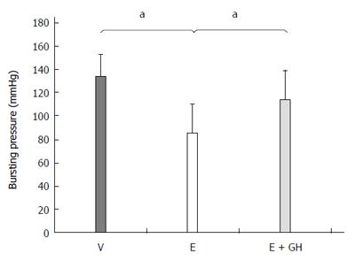
The anastomotic bursting pressure was significantly reduced by everolimus (I: 134 ± 19 mmHg, II: 85 ± 25 mmHg, P < 0.05) (Figure 1). Simultaneous treatment with hGH resulted in a higher bursting pressure (III: 114 ± 25 mmHg). However, this increase was not statistically significantly both compared to groups I and II (P = 0.09, III vs I and III vs II each).

Figure 1
Figure 1 Bursting pressure of the colonic anastomosis (mmHg). Everolimus treatment decreased the bursting pressure significantly. Additional treatment with human growth hormone (hGH) partial antagonized this effect. V: Vehicle, E: Everolimus 3.0 mg/kg, E + GH: Everolimus 3.0 mg/kg + hGH 2.5 mg/kg; aP < 0.05, V vs E, E vs E + GH.
Histology

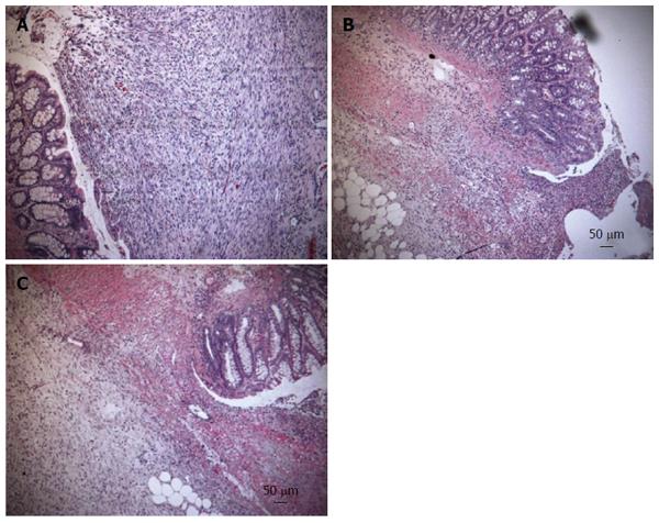
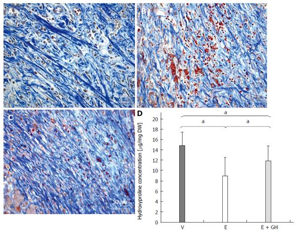
In the H and E stained samples there were significant changes in anastomotical architecture in everolimus-treated animals, which were reduced by simultaneous treatment with hGH (Figure 2A-C). Azan staining revealed a decreased arrangement of the collagen fibres under treatment with only everolimus compared to placebo and simultaneous treatment with hGH (Figure 3A-C).

Figure 2
Figure 2 Hematoxylin and eosin staining from the anastomotic region. In the hematoxylin and eosin stained samples there were significant changes in anastomotical architecture in everolimus-treated animals, indicated by decreased cell density and reduced angiogenesis (B) which are indicators for granulation tissue. Simultaneous treatment with human growth hormone (hGH) showed more granulation tissue (C). A: Vehicle; B: Everolimus 3.0 mg/kg; C: Everolimus 3.0 mg/kg + hGH 2.5 mg/kg (magnification × 100).
Figure 3
Figure 3 Azan staining from the anastomotic region. The collagen fibres appear blue. Everolimus treated animals show a disturbed arrangement of collagen fibres, which is antagonized by additional human growth hormone (hGH)-treatment (A-C). A: Vehicle; B: Everolimus 3.0 mg/kg; C: Everolimus 3.0 mg/kg + hGH 2.5 mg/kg. Hydroxyproline concentration in the anastomotic region in mg per mg dry weight (DW). The decrease of hydroxyproline under everolimus-treatment is reduced by additional hGH-treatment (D) (magnification × 400). V: Vehicle, E: Everolimus 3.0 mg/kg, E + GH: Everolimus 3.0 mg/kg + hGH 2.5 mg/kg; aP < 0.05, vs E group, V vs E + GH group.
Anastomotic hydroxyproline content

Everolimus significantly reduced the hydroxyproline content in the anastomotic region, while simultaneous treatment with hGH inverted this negative effect completely (I: 14.9 ± 2.5 μg/mg DW, II: 8.9 ± 3.6 μg/mg DW, III: 11.9 ± 2.8 μg/mg DW, P < 0.05, I vs II and I vs III and II vs III; Figure 3D).

PCNA and MPO immunohistochemistry

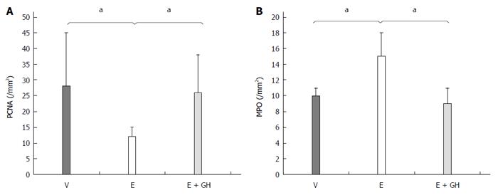
Everolimus decreased PCNA expression significantly compared to placebo treatment (I: 28 ± 3 n/mm², II: 12 ± 3 n/mm², P < 0.05), while simultaneous treatment with hGH antagonized this effect completely (III: 26 ± 12 n/mm², P < 0.05 II vs I and III; Figure 4A). On the other hand hGH lead to normalization of the number of MPO-positive cells compared to only-everolimus-treated animals (I: 10 ± 1 n/mm², II: 15 ± 3 n/mm², III: 9 ± 2 n/mm²; P < 0.05, II vs I and III; Figure 4B).

Figure 4
Figure 4 Immunohistochemistry for PCNA- (A) and MPO- (B) positive cells from the anastomotic region. Treatment with everolimus alone reduced significantly the amount of PCNA-positive cells in the anastomotic region while increasing the number of MPO-positive cells, indicating a prolonged inflammation. Simultaneous treatment with hGH led to nearly normal values for PCNA- and MPO-positive cells. V: Vehicle, E: Everolimus 3.0 mg/kg, E + GH: Everolimus 3.0 mg/kg + hGH 2.5 mg/kg; V vs E, E + GH vs E, aP < 0.05.
Zymography

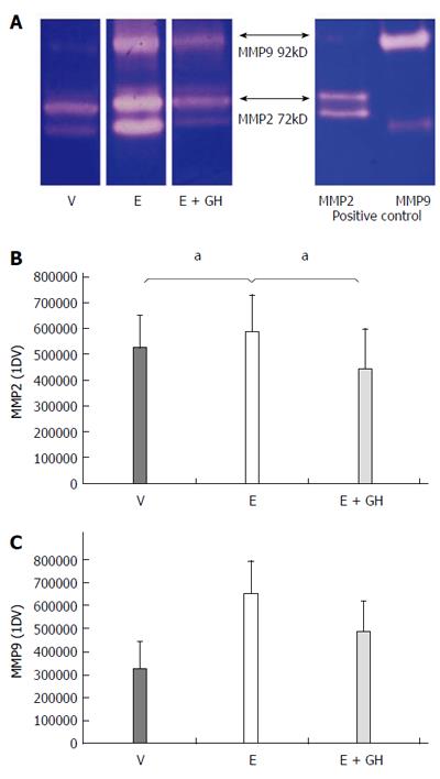
Everolimus-treated animals had significantly increased activity-levels of the pro-inflammatory zytokines MMP-2 and MMP-9 in the anastomotic region. Simultaneous treatment with hGH reduced these activity-levels to nearly-normal values (Figure 5A-C).

Figure 5
Figure 5 Zymographic activities of the pro-inflammatory matrix-metalloproteinases MMP2 and MMP9. Zymography gel showing increased proteolytic activity under treatment with everolimus which is reduced by simultaneous treatment with human growth hormone (hGH) (A). Numeric calculation of the proteolytic activity of MMP2 (B) and MMP9 (C). V: vehicle, E: everolimus 3.0 mg/kg, E+GH: everolimus 3.0 mg/kg + hGH 2.5 mg/kg; V vs E, E + GH vs E, aP < 0.05.
DISCUSSION

Wound healing is a complex biological process which is essential for healing of injured tissue. There are different phases of wound healing finally leading to a stable scar in the wounded tissue. After initial cleaning of the wound by macrophages and neutrophil granulocytes the white cell population shifts to predominantly macrophages, which consume large amounts of oxygen by their respiratory burst which produce proinflammatory cytokines, enzymes (e.g., the matrix-metalloproteinases MMP-2 and MMP-9) and growth factors to remove destroyed collagen from the wounded tissue and to attract cells to the wound which are essential for the proliferation phase[3,43]. This particular step of the healing cascade is defined as the so-called inflammatory phase; its activity peaks in the first 3 to 5 d after wounding[2,44]. Subsequently, healing progresses to the phases of proliferation and tissue remodeling, which are mainly characterized by collagen synthesis by the fibroblasts and new vessel growth to provide enough oxygen to the wound[3,44]. In this phase fibroblasts produce mainly collagen to repair the wound. Moreover, angiogenesis is started to provide enough oxygen to the wound. If these two phases of wound healing are disturbed, it may result in severe wound healing problems which can be devastating in cases of non-healing anastomoses in the gastrointestinal tract. mTOR is a key factor in the regulation of protein synthesis and cell proliferation. Its effects are mediated via complex building with two complexes being involved [mTOR-complex 1 (mTORC1) and mTOR-complex 2 (mTORC2)]. mTORC1 is essential in the regulation of protein synthesis, while the main function of mTORC2 is regulation of the actin cytosceleton. Both, mTORC1 and mTORC2 are activated amongst others by insulin-like growth factor 1 (IGF-1), a growth factor, which is stimulated by the human growth hormone (hGH). IGF-1 acts via the Protein-Kinase B (PKB), also known as Akt, and the corresponding Akt-pathway, resulting in complex-building of mTORC1 and thus activation of protein synthesis. This has been demonstrated in animal studies previously. Christensen et al[34] investigated the effects of hGH on the healing of rodent colon anastomoses in the 1990s[35,45-49]. They found an increased bursting pressure in the hGH-treated animals which showed a significantly increased amount of collagen in the anastomotic region compared to the control groups[35,45,46]. Beckert et al[32] demonstrated in another animal study that administration of hGH leads to increased healing of gastric ulcers in rats compared to the control groups. They showed an increased cell proliferation and angogenesis, both key factors of wound healing[3,44].

On the other hand, mTOR-inhibitors have been demonstrated to significantly disturbe wound healing. Initially there were clinical reports of patients after kidney transplantation who received sirolimus as immunosuppressant drug. These patients had significantly more wound complications than patients with a mTOR-inhibitor-free immunosuppressant regime[21,22,50]. The same was demonstrated later on after liver transplantation[19,20]. van der Vliet et al[27] then showed in 2011 that the hydroxyproline-content in healing colon anastomoses in rats was significantly decreased after treatment with everolimus. With hydroxyproline acting as a progenitor of collagen, this demonstrated for the first time, that mTOR-inihibtors result in a decreased wound strength by reducing protein synthesis. A similar way of action was shown in 2011, when it was demonstrated in the same experimental setting like in this study, that everolimus resulted in a decreased hydroxyproline-content in colonic anastomotic tissue. Furthermore, cell proliferation was reduced as well as angiogenesis while the inflammatory reaction in the anastomosis was increased by means of increased myeloperoxidase (MPO)-positive cells and increased activity of the matrix-metalloproteinases 2 and 9 (MMP2/9) indicating a delayed phase of proliferation[31].

The combination of hGH and everolimus resulted in an improved wound healing compared to everolimus alone. Bursting pressure, content and organization of collagen in the anastomotic tissue, as well as histological parameters like cell density and angiogenesis were significantly improved after combination treatment with hGH and everolimus compared to everolimus alone. All of these factors contribute the proliferation phase of wound healing and were increased, whilst MPO-positive cells and activity of MMP-2 and -9, both markers of the inflammatory phase of wound healing, were reduced by combination treatment of hGH and everolimus. This indicates a partial normalization of wound healing. However, all of these factors were still reduced compared to placebo treatment. One possible explanation is the activation of the Akt-pathway by an IGF-1 synthesis, which is stimulated by hGH. This Akt-pathway leads to an increased forming of the mTOR-complex 1 (mTORC1). On the other hand, everolimus binds to this mTORC1 with high affinity resulting in reduction of the mTORC1-mediated protein-synthesis and cell proliferation. With increased amounts of mTORC1 due to the hGH-treatment, higher doses of everolimus should be needed to produce the same effects as without simultaneous hGH-treatment. However, this has to be investigated in further studies.

In conclusion we could demonstrate for the first time that simultaneous treatment of everolimus and hGH can ameliorate the negative effects of everolimus on intestinal wound healing. However, the exact mechanisms of this partial antagonization remain unclear by now and have to be further investigated.

ACKNOWLEDGMENTS

We thank Dr. Dr. Frank Traub for the histopathological evaluation of the specimen. We thank Prof. Dr. Martin Eichner from the Institute of clinical epidemiology and applied biostatistics from the University of Tübingen for help with the biostatistics. We thank Mrs. Catharina Scheuermann-Poley for language editing.

COMMENTS
Background

Mammalian target of rapamycin (mTOR)-inhibitors are part of standard immunosuppressive medication after both solid organ and bone marrow or stem cell transplantation. The mostly used agents are sirolimus and its derivate everolimus. However, due to severe impairment of wound healing, the definitive mTOR-based immunosuppressive regime is often initiated weeks after transplantation. Moreover, in case of some medical interventions such as surgical procedures the regime has to been switched to an mTOR-inhibitor-free regime with an increased risk of adverse events like graft rejection. To reduce this risk we need to better understand the mechanisms of mTOR-inhibitor-induced impairment of wound healing. One aspect is the delayed inflammatory phase of wound healing resulting in a decreased deposition of wound collagen. So there might be a point of application to reduce this risk.

Research frontiers

The delayed inflammatory phase of wound healing probably is only one mechanism of action of impaired wound healing induced by mTOR-inhibitors as the exact working mechanisms are not known in detail yet. Moreover it is not known yet whether the immunosuppressive potential of the mTOR-inhibitors is caused by the same pathway.

Innovations and breakthroughs

This is the first study which evaluated an approach to counter the negative effects of everolimus on the wound healing and the first time that it was demonstrated that the negative effects of everolimus on wound healing can be antagonized at least partly by another agent.

Applications

The present results might be used to better understand the mechanisms of action of mTOR-inhibitors as well as the basis for further investigation of wound healing under mTOR-inhibitor medication. In the end there is the goal that changing of mTOR-inhibitor-based immunosuppressive regimes is not necessary any more in advance of a planned medical intervention or after an emergency surgical procedure.

Peer-review

The research topic has significant application for immunosuppressant medications in patients undergoing solid organ transplantation

Footnotes

P- Reviewer: Bashashati M, Tran CD S- Editor: Qi Y L- Editor: A E- Editor: Liu XM

References
1.  Coerper S, Beckert S, Becker HD. Possible interventions in impaired wound healing. Chirurg. 2004;75:471-476.  [PubMed]  [DOI]  [Cited in This Article: ]  [Cited by in Crossref: 3]  [Cited by in F6Publishing: 3]  [Article Influence: 0.1]  [Reference Citation Analysis (0)]
2.  Witte MB, Barbul A. General principles of wound healing. Surg Clin North Am. 1997;77:509-528.  [PubMed]  [DOI]  [Cited in This Article: ]  [Cited by in Crossref: 490]  [Cited by in F6Publishing: 439]  [Article Influence: 15.7]  [Reference Citation Analysis (0)]
3.  Hunt TK, Hopf H, Hussain Z. Physiology of wound healing. Adv Skin Wound Care. 2000;13:6-11.  [PubMed]  [DOI]  [Cited in This Article: ]
4.  Broughton G, Janis JE, Attinger CE. The basic science of wound healing. Plast Reconstr Surg. 2006;117:12S-34S.  [PubMed]  [DOI]  [Cited in This Article: ]  [Cited by in Crossref: 854]  [Cited by in F6Publishing: 874]  [Article Influence: 46.0]  [Reference Citation Analysis (0)]
5.  Broughton G, Janis JE, Attinger CE. Wound healing: an overview. Plast Reconstr Surg. 2006;117:1e-S-32e-1e-S-32S.  [PubMed]  [DOI]  [Cited in This Article: ]
6.  Knighton DR, Hunt TK, Thakral KK, Goodson WH. Role of platelets and fibrin in the healing sequence: an in vivo study of angiogenesis and collagen synthesis. Ann Surg. 1982;196:379-388.  [PubMed]  [DOI]  [Cited in This Article: ]  [Cited by in Crossref: 179]  [Cited by in F6Publishing: 168]  [Article Influence: 3.9]  [Reference Citation Analysis (0)]
7.  Thornton FJ, Barbul A. Healing in the gastrointestinal tract. Surg Clin North Am. 1997;77:549-573.  [PubMed]  [DOI]  [Cited in This Article: ]  [Cited by in Crossref: 237]  [Cited by in F6Publishing: 250]  [Article Influence: 8.9]  [Reference Citation Analysis (0)]
8.  Tandara AA, Mustoe TA. The role of the epidermis in the control of scarring: evidence for mechanism of action for silicone gel. J Plast Reconstr Aesthet Surg. 2008;61:1219-1225.  [PubMed]  [DOI]  [Cited in This Article: ]  [Cited by in Crossref: 69]  [Cited by in F6Publishing: 71]  [Article Influence: 4.2]  [Reference Citation Analysis (0)]
9.  Schwentker A, Vodovotz Y, Weller R, Billiar TR. Nitric oxide and wound repair: role of cytokines? Nitric Oxide. 2002;7:1-10.  [PubMed]  [DOI]  [Cited in This Article: ]  [Cited by in Crossref: 262]  [Cited by in F6Publishing: 252]  [Article Influence: 11.0]  [Reference Citation Analysis (0)]
10.  Schwentker A, Billiar TR. Nitric oxide and wound repair. Surg Clin North Am. 2003;83:521-530.  [PubMed]  [DOI]  [Cited in This Article: ]  [Cited by in Crossref: 60]  [Cited by in F6Publishing: 61]  [Article Influence: 2.8]  [Reference Citation Analysis (0)]
11.  Howdieshell TR, Webb WL, Sathyanarayana PL. Inhibition of inducible nitric oxide synthase results in reductions in wound vascular endothelial growth factor expression, granulation tissue formation, and local perfusion. Surgery. 2003;133:528-537.  [PubMed]  [DOI]  [Cited in This Article: ]  [Cited by in Crossref: 29]  [Cited by in F6Publishing: 29]  [Article Influence: 1.3]  [Reference Citation Analysis (0)]
12.  Beckert S, Königsrainer A, Coerper S. The physiology of wound healing. Ther Umsch. 2007;64:467-472.  [PubMed]  [DOI]  [Cited in This Article: ]  [Cited by in Crossref: 2]  [Cited by in F6Publishing: 3]  [Article Influence: 0.2]  [Reference Citation Analysis (0)]
13.  Ortolan EV, Spadella CT, Caramori C, Machado JL, Gregorio EA, Rabello K. Microscopic, morphometric and ultrastructural analysis of anastomotic healing in the intestine of normal and diabetic rats. Exp Clin Endocrinol Diabetes. 2008;116:198-202.  [PubMed]  [DOI]  [Cited in This Article: ]  [Cited by in Crossref: 16]  [Cited by in F6Publishing: 14]  [Article Influence: 0.8]  [Reference Citation Analysis (0)]
14.  Posma LA, Bleichrodt RP, van Goor H, Hendriks T. Transient profound mesenteric ischemia strongly affects the strength of intestinal anastomoses in the rat. Dis Colon Rectum. 2007;50:1070-1079.  [PubMed]  [DOI]  [Cited in This Article: ]  [Cited by in Crossref: 23]  [Cited by in F6Publishing: 26]  [Article Influence: 1.4]  [Reference Citation Analysis (0)]
15.  Stumpf M, Krones CJ, Klinge U, Rosch R, Junge K, Schumpelick V. Collagen in colon disease. Hernia. 2006;10:498-501.  [PubMed]  [DOI]  [Cited in This Article: ]  [Cited by in Crossref: 22]  [Cited by in F6Publishing: 24]  [Article Influence: 1.3]  [Reference Citation Analysis (0)]
16.  Stumpf M, Klinge U, Wilms A, Zabrocki R, Rosch R, Junge K, Krones C, Schumpelick V. Changes of the extracellular matrix as a risk factor for anastomotic leakage after large bowel surgery. Surgery. 2005;137:229-234.  [PubMed]  [DOI]  [Cited in This Article: ]  [Cited by in Crossref: 86]  [Cited by in F6Publishing: 95]  [Article Influence: 4.8]  [Reference Citation Analysis (0)]
17.  Onodera H, Ikeuchi D, Nagayama S, Imamura M. Weakness of anastomotic site in diabetic rats is caused by changes in the integrity of newly formed collagen. Dig Surg. 2004;21:146-151.  [PubMed]  [DOI]  [Cited in This Article: ]  [Cited by in Crossref: 28]  [Cited by in F6Publishing: 30]  [Article Influence: 1.4]  [Reference Citation Analysis (0)]
18.  Flechner SM, Zhou L, Derweesh I, Mastroianni B, Savas K, Goldfarb D, Modlin CS, Krishnamurthi V, Novick A. The impact of sirolimus, mycophenolate mofetil, cyclosporine, azathioprine, and steroids on wound healing in 513 kidney-transplant recipients. Transplantation. 2003;76:1729-1734.  [PubMed]  [DOI]  [Cited in This Article: ]  [Cited by in Crossref: 116]  [Cited by in F6Publishing: 118]  [Article Influence: 5.6]  [Reference Citation Analysis (0)]
19.  Guilbeau JM. Delayed wound healing with sirolimus after liver transplant. Ann Pharmacother. 2002;36:1391-1395.  [PubMed]  [DOI]  [Cited in This Article: ]  [Cited by in Crossref: 71]  [Cited by in F6Publishing: 72]  [Article Influence: 3.1]  [Reference Citation Analysis (0)]
20.  Kahn D, Spearman CW, Mall A, Shepherd E, Engelbrecht G, Lotz Z, Tyler M. Effect of rapamycin on the healing of the bile duct. Transplant Proc. 2005;37:832-833.  [PubMed]  [DOI]  [Cited in This Article: ]  [Cited by in Crossref: 15]  [Cited by in F6Publishing: 15]  [Article Influence: 0.8]  [Reference Citation Analysis (0)]
21.  Kahn D, Spearman CW, Mall A, Shepherd E, Engelbrecht G, Lotz Z, Tyler M. The effect of rapamycin on the healing of the ureteric anastomosis and wound healing. Transplant Proc. 2005;37:830-831.  [PubMed]  [DOI]  [Cited in This Article: ]  [Cited by in Crossref: 20]  [Cited by in F6Publishing: 21]  [Article Influence: 1.1]  [Reference Citation Analysis (0)]
22.  Valente JF, Hricik D, Weigel K, Seaman D, Knauss T, Siegel CT, Bodziak K, Schulak JA. Comparison of sirolimus vs. mycophenolate mofetil on surgical complications and wound healing in adult kidney transplantation. Am J Transplant. 2003;3:1128-1134.  [PubMed]  [DOI]  [Cited in This Article: ]  [Cited by in Crossref: 196]  [Cited by in F6Publishing: 177]  [Article Influence: 8.0]  [Reference Citation Analysis (0)]
23.  Schäffer M, Schier R, Napirei M, Michalski S, Traska T, Viebahn R. Sirolimus impairs wound healing. Langenbecks Arch Surg. 2007;392:297-303.  [PubMed]  [DOI]  [Cited in This Article: ]  [Cited by in Crossref: 37]  [Cited by in F6Publishing: 40]  [Article Influence: 2.2]  [Reference Citation Analysis (0)]
24.  Dean PG, Lund WJ, Larson TS, Prieto M, Nyberg SL, Ishitani MB, Kremers WK, Stegall MD. Wound-healing complications after kidney transplantation: a prospective, randomized comparison of sirolimus and tacrolimus. Transplantation. 2004;77:1555-1561.  [PubMed]  [DOI]  [Cited in This Article: ]  [Cited by in Crossref: 257]  [Cited by in F6Publishing: 260]  [Article Influence: 12.4]  [Reference Citation Analysis (0)]
25.  Dutly AE, Gaspert A, Inci I, Schneiter D, Korom S, Weder W. The influence of the rapamycin-derivate SDZ RAD on the healing of airway anastomoses. Eur J Cardiothorac Surg. 2003;24:154-158; discussion 158.  [PubMed]  [DOI]  [Cited in This Article: ]  [Cited by in Crossref: 20]  [Cited by in F6Publishing: 21]  [Article Influence: 1.0]  [Reference Citation Analysis (0)]
26.  Groetzner J, Kur F, Spelsberg F, Behr J, Frey L, Bittmann I, Vogeser M, Ueberfuhr P, Meiser B, Hatz R. Airway anastomosis complications in de novo lung transplantation with sirolimus-based immunosuppression. J Heart Lung Transplant. 2004;23:632-638.  [PubMed]  [DOI]  [Cited in This Article: ]  [Cited by in Crossref: 119]  [Cited by in F6Publishing: 124]  [Article Influence: 5.9]  [Reference Citation Analysis (0)]
27.  van der Vliet JA, Willems MC, de Man BM, Lomme RM, Hendriks T. Everolimus interferes with healing of experimental intestinal anastomoses. Transplantation. 2006;82:1477-1483.  [PubMed]  [DOI]  [Cited in This Article: ]  [Cited by in Crossref: 29]  [Cited by in F6Publishing: 29]  [Article Influence: 1.6]  [Reference Citation Analysis (0)]
28.  Schäffer M, Fuchs N, Völker J, Schulz T, Kapischke M, Viebahn R. Differential effect of tacrolimus on dermal and intestinal wound healing. J Invest Surg. 2005;18:71-79.  [PubMed]  [DOI]  [Cited in This Article: ]  [Cited by in Crossref: 27]  [Cited by in F6Publishing: 23]  [Article Influence: 1.2]  [Reference Citation Analysis (0)]
29.  Santangelo M, De Rosa P, Spiezia S, Spinosa G, Grassia S, Zuccaro M, Renda A. Healing of surgical incision in kidney transplantation: a single transplant center’s experience. Transplant Proc. 2006;38:1044-1046.  [PubMed]  [DOI]  [Cited in This Article: ]  [Cited by in Crossref: 19]  [Cited by in F6Publishing: 15]  [Article Influence: 0.8]  [Reference Citation Analysis (0)]
30.  Rogers CC, Hanaway M, Alloway RR, Alexander JW, Boardman RE, Trofe J, Gupta M, Merchen T, Buell JF, Cardi M. Corticosteroid avoidance ameliorates lymphocele formation and wound healing complications associated with sirolimus therapy. Transplant Proc. 2005;37:795-797.  [PubMed]  [DOI]  [Cited in This Article: ]  [Cited by in Crossref: 38]  [Cited by in F6Publishing: 38]  [Article Influence: 1.9]  [Reference Citation Analysis (0)]
31.  Küper MA, Schölzl N, Traub F, Mayer P, Weinreich J, Coerper S, Steurer W, Königsrainer A, Beckert S. Everolimus interferes with the inflammatory phase of healing in experimental colonic anastomoses. J Surg Res. 2011;167:158-165.  [PubMed]  [DOI]  [Cited in This Article: ]  [Cited by in Crossref: 15]  [Cited by in F6Publishing: 16]  [Article Influence: 1.0]  [Reference Citation Analysis (0)]
32.  Beckert S, Class N, Farrahi F, Coerper S. Growth hormone enhances gastric ulcer healing in rats. Med Sci Monit. 2004;10:BR255-BR258.  [PubMed]  [DOI]  [Cited in This Article: ]
33.  Tei TM, Kissmeyer-Nielsen P, Flyvbjerg A, Christensen H. Growth hormone is a stimulating but not an essential factor in healing of colon. A study in GH-deficient dwarf rats. Scand J Surg. 2006;95:205-210.  [PubMed]  [DOI]  [Cited in This Article: ]
34.  Christensen H, Chemnitz J, Christensen BC, Oxlund H. Collagen structural organization of healing colonic anastomoses and the effect of growth hormone treatment. Dis Colon Rectum. 1995;38:1200-1205.  [PubMed]  [DOI]  [Cited in This Article: ]  [Cited by in Crossref: 34]  [Cited by in F6Publishing: 34]  [Article Influence: 1.1]  [Reference Citation Analysis (0)]
35.  Christensen H, Oxlund H, Laurberg S. Postoperative biosynthetic human growth hormone increases the strength and collagen deposition of experimental colonic anastomoses. Int J Colorectal Dis. 1991;6:133-138.  [PubMed]  [DOI]  [Cited in This Article: ]  [Cited by in Crossref: 19]  [Cited by in F6Publishing: 20]  [Article Influence: 0.6]  [Reference Citation Analysis (0)]
36.  Petersen TI, Kissmeyer-Nielsen P, Flyvbjerg A, Laurberg S, Christensen H. Effect of insulin-like growth factor I (IGF-I) administration on the healing of colonic anastomoses in rats. Int J Colorectal Dis. 1996;11:19-24.  [PubMed]  [DOI]  [Cited in This Article: ]  [Cited by in Crossref: 18]  [Cited by in F6Publishing: 18]  [Article Influence: 0.6]  [Reference Citation Analysis (0)]
37.  Yarimkaya A, Apaydin B, Unal E, Karabicak I, Aydogan F, Uslu E, Erginoz E, Artis T, Eyuboglu E. Effects of recombinant human growth hormone and nandrolone phenylpropionate on the healing of ischemic colon anastomosis in rats. Dis Colon Rectum. 2003;46:1690-1697.  [PubMed]  [DOI]  [Cited in This Article: ]  [Cited by in Crossref: 14]  [Cited by in F6Publishing: 15]  [Article Influence: 0.7]  [Reference Citation Analysis (0)]
38.  Egger B, Tolmos J, Procaccino F, Sarosi I, Friess H, Büchler MW, Stamos M, Eysselein VE. Keratinocyte growth factor promotes healing of left-sided colon anastomoses. Am J Surg. 1998;176:18-24.  [PubMed]  [DOI]  [Cited in This Article: ]  [Cited by in Crossref: 39]  [Cited by in F6Publishing: 40]  [Article Influence: 1.5]  [Reference Citation Analysis (0)]
39.  Agren MS, Andersen TL, Mirastschijski U, Syk I, Schiødt CB, Surve V, Lindebjerg J, Delaissé JM. Action of matrix metalloproteinases at restricted sites in colon anastomosis repair: an immunohistochemical and biochemical study. Surgery. 2006;140:72-82.  [PubMed]  [DOI]  [Cited in This Article: ]  [Cited by in Crossref: 55]  [Cited by in F6Publishing: 60]  [Article Influence: 3.2]  [Reference Citation Analysis (0)]
40.  Stegemann H, Stalder K. Determination of hydroxyproline. Clin Chim Acta. 1967;18:267-273.  [PubMed]  [DOI]  [Cited in This Article: ]  [Cited by in Crossref: 1202]  [Cited by in F6Publishing: 1203]  [Article Influence: 20.7]  [Reference Citation Analysis (0)]
41.  Beckert S, Hierlemann H, Müschenborn N, Witte M, Ranke M, Coerper S. Experimental ischemic wounds: correlation of cell proliferation and insulin-like growth factor I expression and its modification by different local IGF-I release systems. Wound Repair Regen. 2005;13:278-283.  [PubMed]  [DOI]  [Cited in This Article: ]  [Cited by in Crossref: 20]  [Cited by in F6Publishing: 20]  [Article Influence: 1.0]  [Reference Citation Analysis (0)]
42.  Narita T, Akiyama M. Collagenous colitis in a Japanese patient. Pathol Int. 1996;46:211-215.  [PubMed]  [DOI]  [Cited in This Article: ]  [Cited by in Crossref: 5]  [Cited by in F6Publishing: 5]  [Article Influence: 0.2]  [Reference Citation Analysis (0)]
43.  Weiss SJ. Tissue destruction by neutrophils. N Engl J Med. 1989;320:365-376.  [PubMed]  [DOI]  [Cited in This Article: ]  [Cited by in Crossref: 3068]  [Cited by in F6Publishing: 2950]  [Article Influence: 81.9]  [Reference Citation Analysis (0)]
44.  Witte MB, Barbul A. Repair of full-thickness bowel injury. Crit Care Med. 2003;31:S538-S546.  [PubMed]  [DOI]  [Cited in This Article: ]  [Cited by in Crossref: 45]  [Cited by in F6Publishing: 43]  [Article Influence: 2.0]  [Reference Citation Analysis (0)]
45.  Christensen H, Oxlund H, Laurberg S. Growth hormone increases the bursting strength of colonic anastomoses. An experimental study in the rat. Int J Colorectal Dis. 1990;5:130-134.  [PubMed]  [DOI]  [Cited in This Article: ]  [Cited by in Crossref: 22]  [Cited by in F6Publishing: 23]  [Article Influence: 0.7]  [Reference Citation Analysis (0)]
46.  Christensen H, Jørgensen PH, Oxlund H, Laurberg S. Growth hormone increases the mass, the collagenous proteins, and the strength of rat colon. Scand J Gastroenterol. 1990;25:1137-1143.  [PubMed]  [DOI]  [Cited in This Article: ]  [Cited by in Crossref: 17]  [Cited by in F6Publishing: 18]  [Article Influence: 0.5]  [Reference Citation Analysis (0)]
47.  Christensen H, Oxlund H. Growth hormone increases the collagen deposition rate and breaking strength of left colonic anastomoses in rats. Surgery. 1994;116:550-556.  [PubMed]  [DOI]  [Cited in This Article: ]
48.  Christensen H, Flyvbjerg A. Dose-dependent stimulatory effect of human growth hormone on the strength and collagen deposition of colonic anastomoses in the rat. Acta Endocrinol (Copenh). 1992;126:438-443.  [PubMed]  [DOI]  [Cited in This Article: ]  [Cited by in Crossref: 15]  [Cited by in F6Publishing: 15]  [Article Influence: 0.5]  [Reference Citation Analysis (0)]
49.  Christensen H, Andreassen TT, Oxlund H. Increased mechanical strength of left colon in old rats treated with growth hormone. Gerontology. 1992;38:245-251.  [PubMed]  [DOI]  [Cited in This Article: ]  [Cited by in Crossref: 11]  [Cited by in F6Publishing: 11]  [Article Influence: 0.3]  [Reference Citation Analysis (0)]
50.  Troppmann C, Pierce JL, Gandhi MM, Gallay BJ, McVicar JP, Perez RV. Higher surgical wound complication rates with sirolimus immunosuppression after kidney transplantation: a matched-pair pilot study. Transplantation. 2003;76:426-429.  [PubMed]  [DOI]  [Cited in This Article: ]  [Cited by in Crossref: 111]  [Cited by in F6Publishing: 109]  [Article Influence: 5.0]  [Reference Citation Analysis (0)]