Published online Aug 28, 2005. doi: 10.3748/wjg.v11.i32.5079
Revised: November 23, 2004
Accepted: December 1, 2004
Published online: August 28, 2005
Schwannomas are rare tumors derived from the cells of Schwann that form the neural sheath. When located in the gastrointestinal tract, they constitute together with leiomyoma, leiomyoblastoma, and leiomyosarcoma, the gastrointestinal stromal tumors (GIST). Peripheral nerve sheath tumors represent 2-6% GIST with most common location, the stomach and the small intestine. Schwannomas of the colon and rectum are extremely rare and radical excision with wide margins is mandatory, due to their tendency to recur locally and become malignant, if left untreated. In the present study, we report a rare case of a sigmoid schwannoma, which was successfully treated in our department and reviewed the literature.
- Citation: Fotiadis CI, Kouerinis IA, Papandreou I, Zografos GC, Agapitos G. Sigmoid schwannoma: A rare case. World J Gastroenterol 2005; 11(32): 5079-5081
- URL: https://www.wjgnet.com/1007-9327/full/v11/i32/5079.htm
- DOI: https://dx.doi.org/10.3748/wjg.v11.i32.5079
Gastrointestinal stromal tumors (GIST) include a variety of primary mesenchymal tumors of the gastrointestinal tract such as leiomyoma, leiomyosarcoma, leiomyoblastoma, and schwannoma[1]. They have recently been distinguished from each other on the basis of the tumor cell differentiation revealed by immunohistochemical studies[2,3]. Besides the above-mentioned pathologies, the term GIST has been recently applied to mesenchymal tumors that represent neither typical leiomyoma nor schwannoma[2,4].
It is well established that schwannomas appear more frequently in the stomach and the small intestine, while location in the colon or in the rectum is uncommon[5,6]. Most of them are benign and asymptomatic; nevertheless, the possibility of malignant degeneration does exist and is directly related to the tumor dimensions. Radical surgical treatment is the gold standard in all cases, since the results of both chemotherapy and radiotherapy remain uncertain[7].
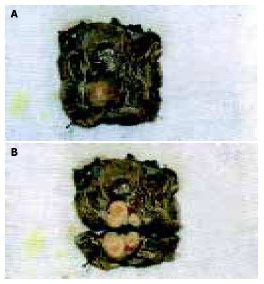
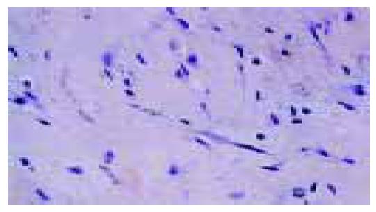
A 55-year-old male patient was admitted to our department, with a chief complaint of intermittent rectal bleeding and diarrhea that had started 2 mo before admission. Physical findings and digital examination were undiagnostic, but laboratory tests disclosed red blood cell count: 3.2×10⁵/mm3, hemoglobin: 74 g/L, and hematocrit: 26.4%. Colonoscopy revealed a wide-based mass protruding from the posterior wall of sigmoid and multiple biopsies were taken from different parts of the tumor. Abdominal x-rays were negative for bowel obstruction, while CT scans proved no metastatic disease. Additionally, the serum levels of tumor markers including carcinoembryonic antigen and Ca 19-9 were within normal ranges. The diagnosis of schwannoma Antoni B type was set preoperatively based on the endoscopic biopsies and intraoperatively, segmental resection of the tumor with wide margins was performed (Figures 1A and B). Permanent sections with H&E confirmed the preoperative diagnosis (Figure 2). The patient recovered uneventfully and at 5 years following surgery, he was free of disease without receiving adjuvant therapy.
Schwannomas are mainly benign tumors derived from the cells of Schwann that form the neural sheath, which may become malignant, if left untreated[8-10]. They can be located everywhere, but the most common type of benign schwannoma is the acoustic neuroma (VIII cranial nerve)[11].
Malignant schwannomas usually come under the heading of soft tissue sarcomas and the gastrointestinal tract is one of the sites that such tumors can be found[12]. In these cases they constitute together with leiomyoma, leiomyosarcoma, leiomyoblastoma, and another type with mixed characteristics between leiomyoma and schwannoma, the GIST[13]. All of these primary mesenchymal tumors show a wide range of common histological and immunohistochemical characteristics[2,14].
Peripheral nerve sheath tumors represent 2-6% of stromal tumors of the gastrointestinal tract[3,4] but reports of solitary schwannomas of the rectum and colon are even more rare[15]. They have almost the same incidence in men and women and a median age of presentation, 65 years of age[4]. Their size may vary; a case of a rectal schwannoma with a diameter of 8 cm which was primarily misdiagnosed as a myogenic sarcoma has been reported[2,5].
Although, they usually grow very slowly and are asymptomatic, sometimes they may cause rectal bleeding, colonic obstruction, defecation disorders, and pain[3,4]. It is very important to emphasize that rectal pain can also be the only symptom of intraspinal schwannomas which they often mislead the diagnosis to proctalgia fugax or rectal neuralgia[15].
Two histologic types of schwannomas have been described; Antoni type A with densely packed spindle cells (Verocay bodies) and Antoni type B with loosely organized spindle cells (absence of Verocay bodies) in myxoid stroma[3,4]. Imm-unology plays a central role to the diagnosis of schwannoma as in many other types of cancer[16]. Strongly positive S-100 protein, low affinity, nerve growth factor receptor (P75), collagen IV, GFAP, CD34, and negative CD117, neurof-ilament protein, smooth muscle actin, and desmin, turn out to be supportive to the diagnosis of the benign tumor as a schwannoma[2,3,12]. Schwannomas may also appear in patients with Von Recklinghausen disease (NF I), although there is no evident connection between colorectal schwannomas and NF I[17].
Radical excision with margins free of disease is the treatment of choice, since their response to both chemotherapy and radiotherapy remain uncertain[4,18]. Nevertheless, despite aggressive surgical management, these tumors appear with a high rate of local recurrence and malignant degeneration. In such case’s the treatment options are few and the prognosis is poor[3,7,8].
The surgical approach depends on the tumor size and histologic features that mainly determine the prognosis. Preoperative imaging tests including abdominal x-rays, transanal 3D-ECHO, MRI, and CT scans can be very useful in the selection of patients for surgical treatment[19]. Besides the commonly used surgical techniques, minimally invasive modalities can be performed too. In the transanal endoscopic microsurgery (TEM), the local excision is accomplished through a rectal expander[20,21] with or without the use of an ultrasonically activated scalpel[22]. Alternatively, in cases of the benign schwannomas located on the distal parts of the rectum, a transanal resection can be performed followed by recto-anal anastomosis[21].
In conclusion, rectal schwannoma is a very rare tumor, whose diagnostic approach and treatment present certain gray areas. Nevertheless, it has become obvious that the preoperative histological identification of the tumor followed by a radical surgical resection is the cornerstone, which will determine the overall outcome.
Science Editor Wang XL and Guo SY Language Editor Elsevier HK
1. | Prévot S, Bienvenu L, Vaillant JC, de Saint-Maur PP. Benign schwannoma of the digestive tract: a clinicopathologic and immunohistochemical study of five cases, including a case of esophageal tumor. Am J Surg Pathol. 1999;23:431-436. [PubMed] [DOI] [Cited in This Article: ] [Cited by in Crossref: 113] [Cited by in F6Publishing: 113] [Article Influence: 4.3] [Reference Citation Analysis (0)] |
2. | Miettinen M, Virolainen M. Gastrointestinal stromal tumors--value of CD34 antigen in their identification and separation from true leiomyomas and schwannomas. Am J Surg Pathol. 1995;19:207-216. [PubMed] [DOI] [Cited in This Article: ] [Cited by in Crossref: 327] [Cited by in F6Publishing: 341] [Article Influence: 11.4] [Reference Citation Analysis (0)] |
3. | Miettinen M, Shekitka KM, Sobin LH. Schwannomas in the colon and rectum: a clinicopathologic and immunohistochemical study of 20 cases. Am J Surg Pathol. 2001;25:846-855. [PubMed] [DOI] [Cited in This Article: ] [Cited by in Crossref: 181] [Cited by in F6Publishing: 149] [Article Influence: 6.2] [Reference Citation Analysis (0)] |
4. | Miettinen M, Sarlomo-Rikala M, Lasota J. Gastrointestinal stromal tumours. Ann Chir Gynaecol. 1998;87:278-281. [PubMed] [Cited in This Article: ] |
5. | Maciejewski A, Lange D, Włoch J. Case report of schwannoma of the rectum--clinical and pathological contribution. Med Sci Monit. 2000;6:779-782. [PubMed] [Cited in This Article: ] |
6. | Bhardwaj K, Bal MS, Kumar P. Rectal schwannoma. Indian J Gastroenterol. 2002;21:116-117. [PubMed] [Cited in This Article: ] |
7. | Arcidiaco M, Uggeri G, Rumi A, Valenti L. Schwannoma of the rectum. Clinico-therapeutic study. Minerva Chir. 1981;36:267-272. [PubMed] [Cited in This Article: ] |
8. | Catania G, Puleo C, Cardì F, Catalano F, Iuppa A, Buffone A. Malignant schwannoma of the rectum: a clinical and pathological contribution. Chir Ital. 2001;53:873-877. [PubMed] [Cited in This Article: ] |
9. | Kołodziejski LS, Dyczek ST, Pogodzinski M. Surgical management of retrorectal tumors. J Chir (Paris). 2004;141:109-113. [PubMed] [Cited in This Article: ] |
10. | Reinbold WD, Hillemanns A, Seesko H, Jehn E. Malignant schwannoma of the rectum. Radiologe. 1996;36:663-666. [PubMed] [DOI] [Cited in This Article: ] [Cited by in Crossref: 4] [Cited by in F6Publishing: 5] [Article Influence: 0.2] [Reference Citation Analysis (0)] |
11. | Darrouzet V, Martel J, Enée V, Bébéar JP, Guérin J. Vestibular schwannoma surgery outcomes: our multidisciplinary experience in 400 cases over 17 years. Laryngoscope. 2004;114:681-688. [PubMed] [DOI] [Cited in This Article: ] [Cited by in Crossref: 147] [Cited by in F6Publishing: 145] [Article Influence: 6.9] [Reference Citation Analysis (0)] |
12. | Daimaru Y, Kido H, Hashimoto H, Enjoji M. Benign schwannoma of the gastrointestinal tract: a clinicopathologic and immunohistochemical study. Hum Pathol. 1988;19:257-264. [PubMed] [DOI] [Cited in This Article: ] [Cited by in Crossref: 220] [Cited by in F6Publishing: 205] [Article Influence: 5.5] [Reference Citation Analysis (0)] |
13. | Shibata Y, Ueda T, Seki H, Yagihashi N. Gastrointestinal stromal tumour of the rectum. Eur J Gastroenterol Hepatol. 2001;13:283-286. [PubMed] [DOI] [Cited in This Article: ] [Cited by in Crossref: 19] [Cited by in F6Publishing: 21] [Article Influence: 0.9] [Reference Citation Analysis (0)] |
14. | Vorobév GI, Odariuk TS, Shelygin IuA, Kapuller LL, Korniak BS, Tikhonov AA, Orlova LP. Differential diagnosis of non-epithelial rectal neoplasms. Khirurgiia (Mosk). 1995;45-50. [PubMed] [Cited in This Article: ] |
15. | Kornel EE, Vlahakos D. Intraspinal schwannoma presenting solely with rectal pain. Neurosurgery. 1988;22:417-419. [PubMed] [DOI] [Cited in This Article: ] [Cited by in Crossref: 4] [Cited by in F6Publishing: 6] [Article Influence: 0.2] [Reference Citation Analysis (0)] |
16. | Kouerinis IA, Zografos G, Tarassi KE, Athanasiades TH, Liontos M, Gorgoulis VG, Korkolis D, Konstandoulakis MM, Fotiadis CI, Androulakis G. Human leukocyte antigens as genetic markers in Greek patients with sporadic pancreatic cancer. Pancreas. 2004;29:41-44. [PubMed] [DOI] [Cited in This Article: ] [Cited by in Crossref: 3] [Cited by in F6Publishing: 3] [Article Influence: 0.1] [Reference Citation Analysis (0)] |
17. | Genna M, Leopardi F, Fambri P, Postorino A. Neurogenic tumors of the ano-rectal region. Ann Ital Chir. 1997;68:351-353; discussion 351-353;. [PubMed] [Cited in This Article: ] |
18. | Pollock J, Morgan D, Denobile J, Williams J. Adjuvant radiotherapy for gastrointestinal stromal tumor of the rectum. Dig Dis Sci. 2001;46:268-272. [PubMed] [DOI] [Cited in This Article: ] [Cited by in Crossref: 25] [Cited by in F6Publishing: 27] [Article Influence: 1.1] [Reference Citation Analysis (0)] |
19. | Stroh C, Manger T. Ultrasound diagnosis of rare retrorectal tumors. Zentralbl Chir. 2003;128:1075-1079. [PubMed] [Cited in This Article: ] |
20. | Kakizoe S, Kuwahara S, Kakizoe K, Kakizoe H, Kakizoe Y, Kakizoe T, Yamamoto O, Kakizoe S. Local excision of benign rectal schwannoma using rectal expander-assisted transanal endoscopic microsurgery. Gastrointest Endosc. 1998;48:90-92. [PubMed] [DOI] [Cited in This Article: ] [Cited by in Crossref: 8] [Cited by in F6Publishing: 9] [Article Influence: 0.3] [Reference Citation Analysis (0)] |
21. | Pokrovskiĭ GA, Eropkin PV, Shelygin IuA, Peresada IV. Transanal resection of the rectum. Khirurgiia (Mosk). 1994;32-34. [PubMed] [Cited in This Article: ] |
22. | Langer C, Markus P, Liersch T, Füzesi L, Becker H. UltraCision or high-frequency knife in transanal endoscopic microsurgery (TEM)? Advantages of a new procedure. Surg Endosc. 2001;15:513-517. [PubMed] [DOI] [Cited in This Article: ] [Cited by in Crossref: 42] [Cited by in F6Publishing: 31] [Article Influence: 1.3] [Reference Citation Analysis (0)] |