Copyright
©The Author(s) 2003.
World J Gastroenterol. Sep 15, 2003; 9(9): 1904-1908
Published online Sep 15, 2003. doi: 10.3748/wjg.v9.i9.1904
Published online Sep 15, 2003. doi: 10.3748/wjg.v9.i9.1904
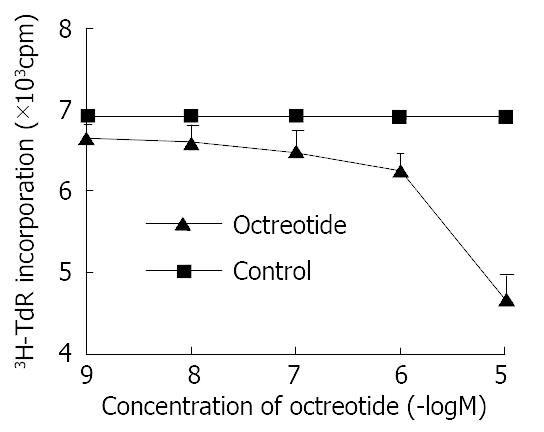
Figure 1 Effects of octreotide on 3H-thymidine incorporation of SGC-7901 cell.
Each value was the mean ± SD of three sepa-rate experiments in which duplicate determinations were made.
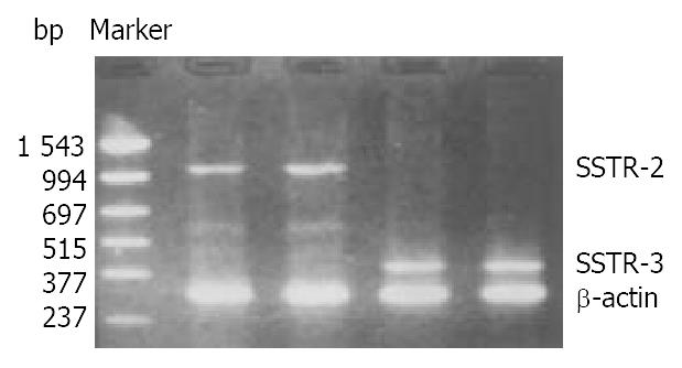
Figure 2 Expression of mRNA for SSTR-2 and SSTR-3 in SGC-7901 cells.
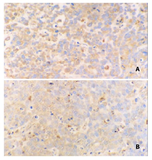
Figure 3 Immunohistochemical staining for ERK in tissues of transplanted gastric cancer in nude mice.
A. control, B. octreotide group (200 ×).
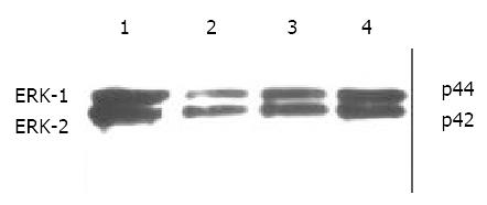
Figure 4 Western blot analysis of ERK in SGC-7901 cells.
1. control, 2. octreotide 1 × 10⁻⁵ M, 3. octreotide 1 × 10⁻⁶ M, 4. octreotide 1 × 10⁻⁷ M.
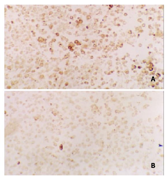
Figure 5 Immunohistochemical staining for c-Fos in tissues of transplanted gastric cancer in nude mice.
A. control, B. ctreotide group (200 ×).
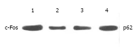
Figure 6 Western blot analysis of c-Fos in SGC-7901 cells.
1. control, 2. Octreotide 1 × 10⁻⁵ M, 3. Octreotide 1 × 10⁻⁶ M, 4. Octreotide 1 × 10⁻⁷ M.
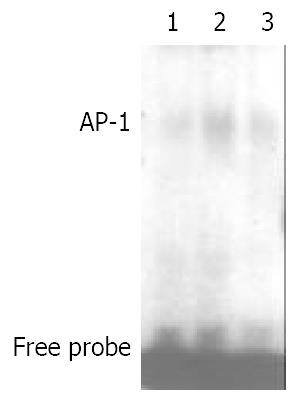
Figure 7 Effects of octreotide on AP-1 binding activity of SGC-7901 cells.
1. control, 2. FCS stimulated, 3. FCS stimulated and octreotide.
-
Citation: Wang CH, Tang CW, Liu CL, Tang LP. Inhibitory effect of octreotide on gastric cancer growth
via MAPK pathway. World J Gastroenterol 2003; 9(9): 1904-1908 - URL: https://www.wjgnet.com/1007-9327/full/v9/i9/1904.htm
- DOI: https://dx.doi.org/10.3748/wjg.v9.i9.1904


