Copyright
©The Author(s) 2003.
World J Gastroenterol. Nov 15, 2003; 9(11): 2596-2600
Published online Nov 15, 2003. doi: 10.3748/wjg.v9.i11.2596
Published online Nov 15, 2003. doi: 10.3748/wjg.v9.i11.2596
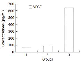
Figure 1 Comparison of VEGF concentrations in different kinds of ascites.
Group 1: cirrhotic ascites, Group 2: tuberculous ascites, Group 3: malignant ascites.
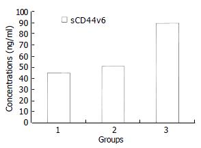
Figure 2 Comparison of sCD44v6 concentrations in different kinds of ascites.
Group 1: cirrhotic ascites, Group 2: tuberculous ascites, Group 3: malignant ascites. Concentrations of sCD44v6 in group 3 were significantly higher than those in groups 1 and 2 (P < 0.01).
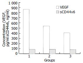
Figure 3 Concentrations of VEGF and sCD44v6 in different kinds of malignant ascites.
Group A: ovarian cancer; Group B: gastric cancer; Group C: colon cancer. Concentrations of VEGF in group A were higher than those in groups B and C (P < 0.01), while the difference of CD44v6 levels among groups A, B and C was not statistically significant (P > 0.05).
- Citation: Dong WG, Sun XM, Yu BP, Luo HS, Yu JP. Role of VEGF and CD44v6 in differentiating benign from malignant ascites. World J Gastroenterol 2003; 9(11): 2596-2600
- URL: https://www.wjgnet.com/1007-9327/full/v9/i11/2596.htm
- DOI: https://dx.doi.org/10.3748/wjg.v9.i11.2596


