Copyright
©The Author(s) 2000.
World J Gastroenterol. Dec 15, 2000; 6(6): 833-841
Published online Dec 15, 2000. doi: 10.3748/wjg.v6.i6.833
Published online Dec 15, 2000. doi: 10.3748/wjg.v6.i6.833
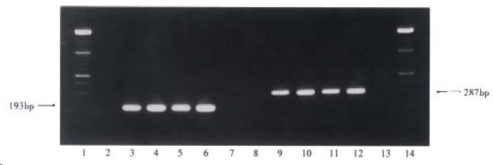
Figure 1 Target amplification fragments from GBV-C 5’-NCR and NS3 region.
(1 and 14: markers; 2 and 8: negative serum samples; 7 and 13: blanks; 3-6: four GBV-C 5’-NCR RNA positive serum samples; 9-12: four GBV-C NS3 region RNA positive serum samples)
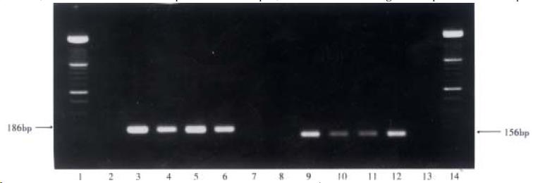
Figure 2 Target amplification fragments from HGV 5’-NCR and NS5 region.
(1 and 14: markers; 2 and 8: negative serum samples; 7 and 13: blanks; 3-6: four HGV 5’-NCR RNA positive serum samples; 9-12: four HGV NS5 region RNA positive serum samples)
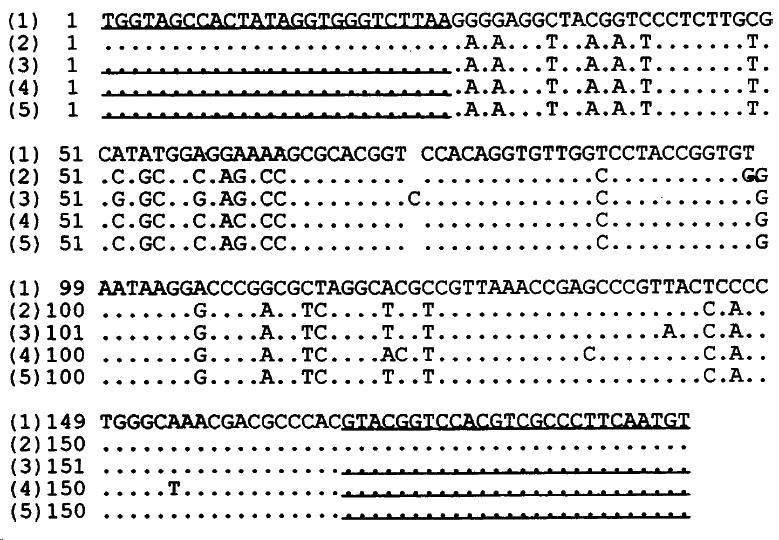
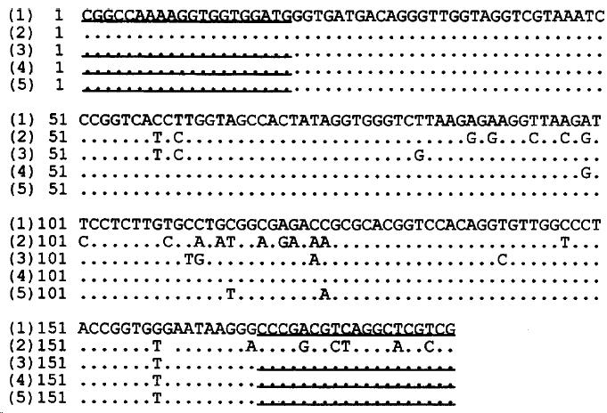
Figure 3 Homology of the nucleotide sequences from GBV-C 5’-NCR RT-nested PCR products from 3 serum samples as compared with the reported sequences.
(1) the reported GBV-C 5’-NCR sequence, (2) the reported HGV 5’-NCR sequence, (3)-(5) the sequences of GBV-C 5’-NCR RT-nested PCR product from 3 samples. Underlined areas indicated the primers’position.
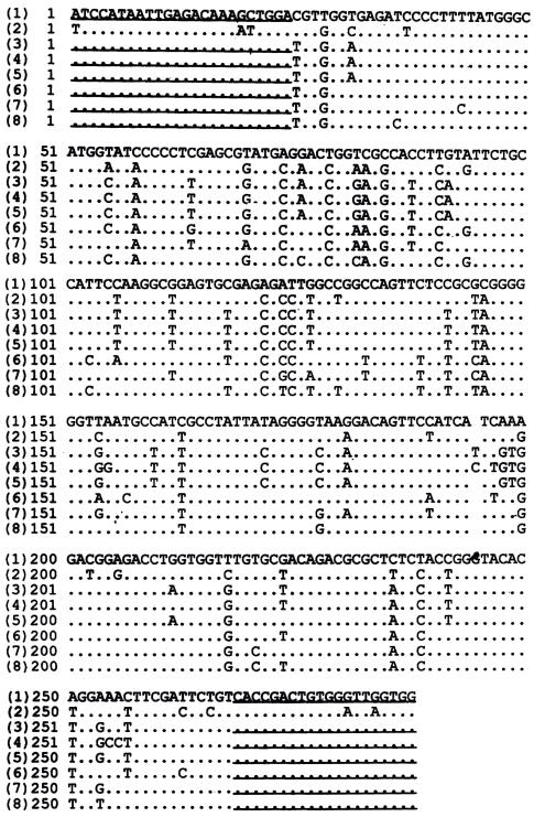
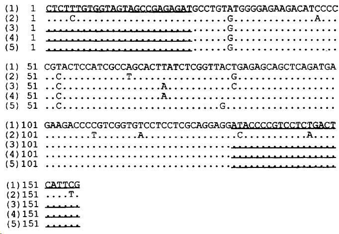
Figure 4 Homology of the sequences from GBV-C NS3 RT-nested PCR products from 6 serum samples compared with the reported sequences.
(1) the reported GBV-C NS3 region sequence, (2) the reported HGV NS3 region sequence, (3)-(8) the sequences from GBV-C NS3 region RT-nested PCR products from 6 serum samples. Underlined areas indicated the primers’position.
Figure 5 Homology of the nucleotide sequences of HGV 5’-NCR RT-PCR products from 3 serum samples compared to the reported sequences.
(1) the reported HGV 5’-NCR sequence, (2) the reported GBV-C 5’-NCR sequence, (3)-(5) the sequences of HGV 5’-NCR RT-PCR products from 3 samples. Underlined indicated the primers’ position.
Figure 6 Homology of the nucleotide sequences from HGV NS5 RT-PCR products from 3 serum samples compared to the reported sequences.
(1) the reported HGV NS5 region sequence, (2) the reported GBV-C NS5 region sequence, (3)-(5) the sequences from HGV NS5 region RT-PCR products from 3 serum samples. Underlined areas indicated the primers’ position.
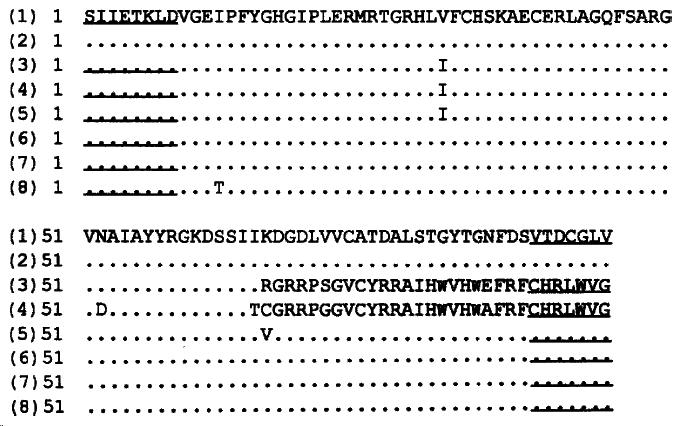
Figure 7 Comparison of the putative amino acid sequences from GBV-C NS3 RT-nested PCR products from 9 serum samples with the reported sequences.
(1) the reported GBV-C sequence, (2) the reported HGV sequence, (3)-(8) putative amino acid sequences translated from the 6 nucleotide sequences from GBV-C NS3 region. Underlined are indicated the primers’ position.
Figure 8 Comparison of the putative amino acid sequences from HGV NS5 RT-PCR products from 3 serum samples with the reported sequences.
(1) the reported HGV sequence, (2) the reported GBV-C sequence, (3)-(5) putative amino acid sequences translated from the 3 nucleotide sequences from HGV NS5 region. Underlined areas indicated the primers’ position.
- Citation: Yan J, Dennin RH. A high frequency of GBV-C/HGV coinfection in hepatitis C patients in Germany. World J Gastroenterol 2000; 6(6): 833-841
- URL: https://www.wjgnet.com/1007-9327/full/v6/i6/833.htm
- DOI: https://dx.doi.org/10.3748/wjg.v6.i6.833


