Copyright
©The Author(s) 2023.
World J Gastroenterol. Mar 28, 2023; 29(12): 1899-1910
Published online Mar 28, 2023. doi: 10.3748/wjg.v29.i12.1899
Published online Mar 28, 2023. doi: 10.3748/wjg.v29.i12.1899
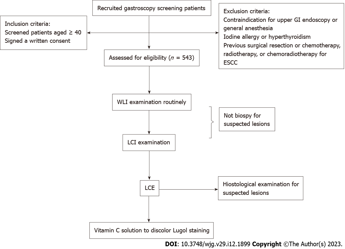
Figure 1 Flow chart of study design.
ESCC: Esophageal squamous cell carcinoma; GI: Gastrointestinal; LCE: Lugol chromoendoscopy; LCI: Linked color imaging; WLI: White light imaging.
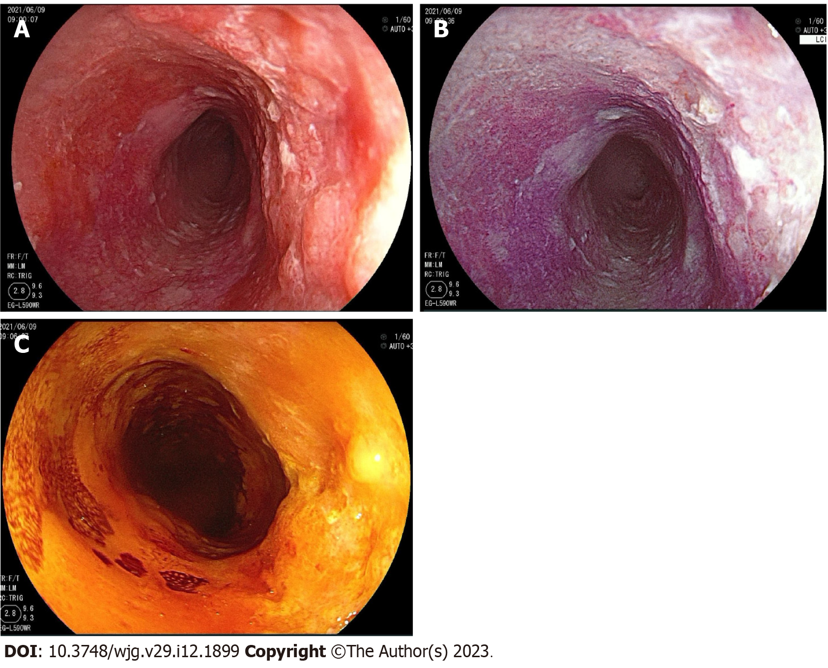
Figure 2 A single esophageal squamous cell carcinoma lesion visualized under the three detection methods.
A: Under white light imaging; B: Under linked color imaging; C: Under Lugol chromoendoscopy.
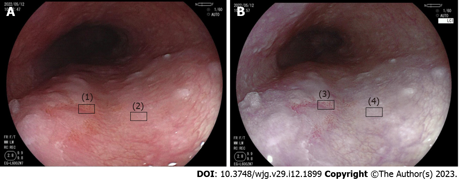
Figure 3 Method to measure the color difference between neoplastic and non-neoplastic lesions detected by white light imaging and linked color imaging.
A: Neoplastic lesion (1) and non-neoplastic lesion (2) detected by white light imaging; B: Neoplastic lesion (3) and non-neoplastic lesion (4) detected by linked color imaging.
- Citation: Wang ZX, Li LS, Su S, Li JP, Zhang B, Wang NJ, Liu SZ, Wang SS, Zhang S, Bi YW, Gao F, Shao Q, Xu N, Shao BZ, Yao Y, Liu F, Linghu EQ, Chai NL. Linked color imaging vs Lugol chromoendoscopy for esophageal squamous cell cancer and precancerous lesion screening: A noninferiority study. World J Gastroenterol 2023; 29(12): 1899-1910
- URL: https://www.wjgnet.com/1007-9327/full/v29/i12/1899.htm
- DOI: https://dx.doi.org/10.3748/wjg.v29.i12.1899


