Copyright
©The Author(s) 2021.
World J Gastroenterol. Feb 7, 2021; 27(5): 442-448
Published online Feb 7, 2021. doi: 10.3748/wjg.v27.i5.442
Published online Feb 7, 2021. doi: 10.3748/wjg.v27.i5.442
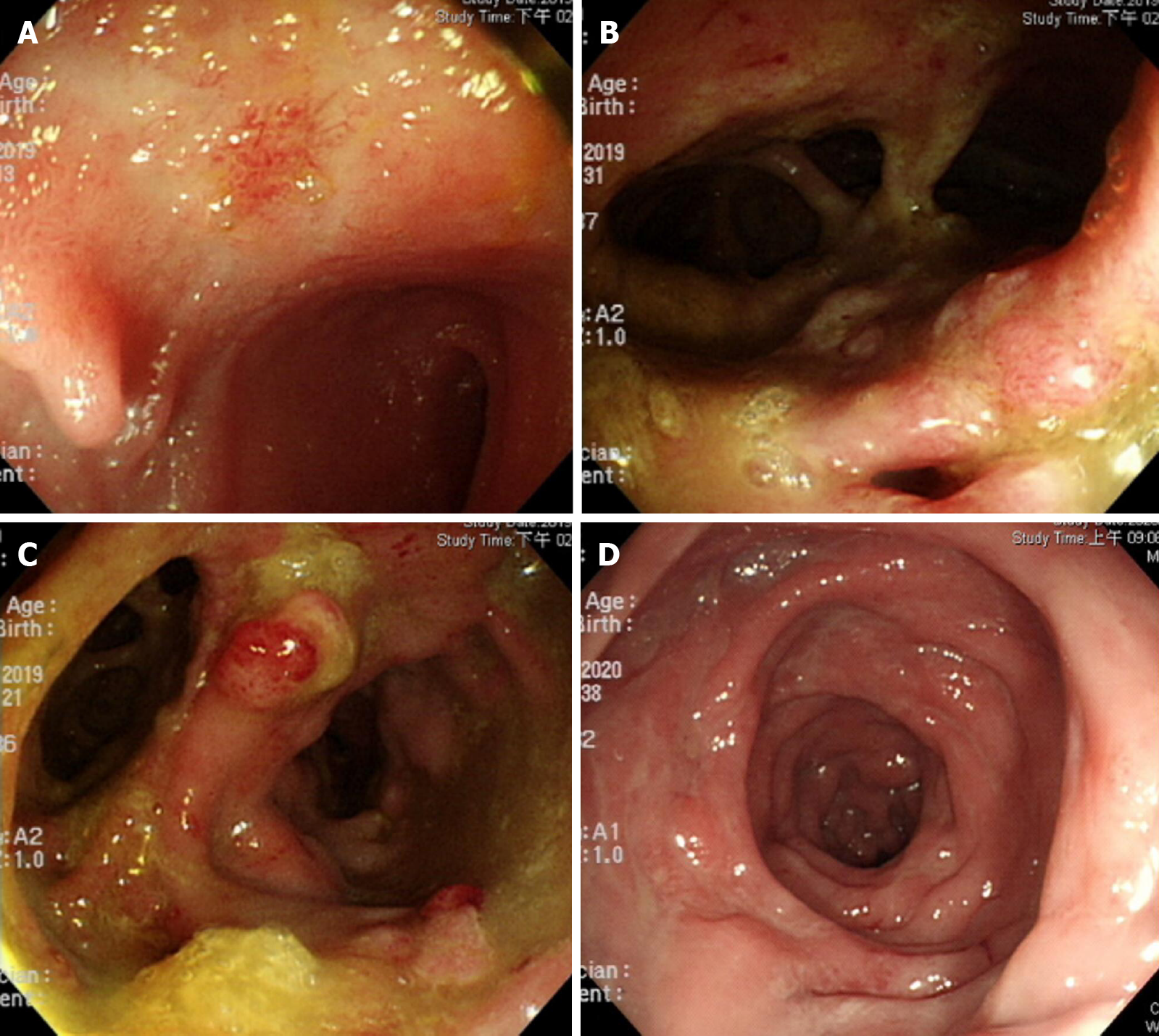
Figure 1 Endoscopic findings.
A: Terminal ileal shallow ulcer at diagnosis; B and C: Multiple rectal fistula tracts with inflammation; D: Mucosal healing without fistula tracts six months after vedolizumab treatment, seven months after diagnosis.
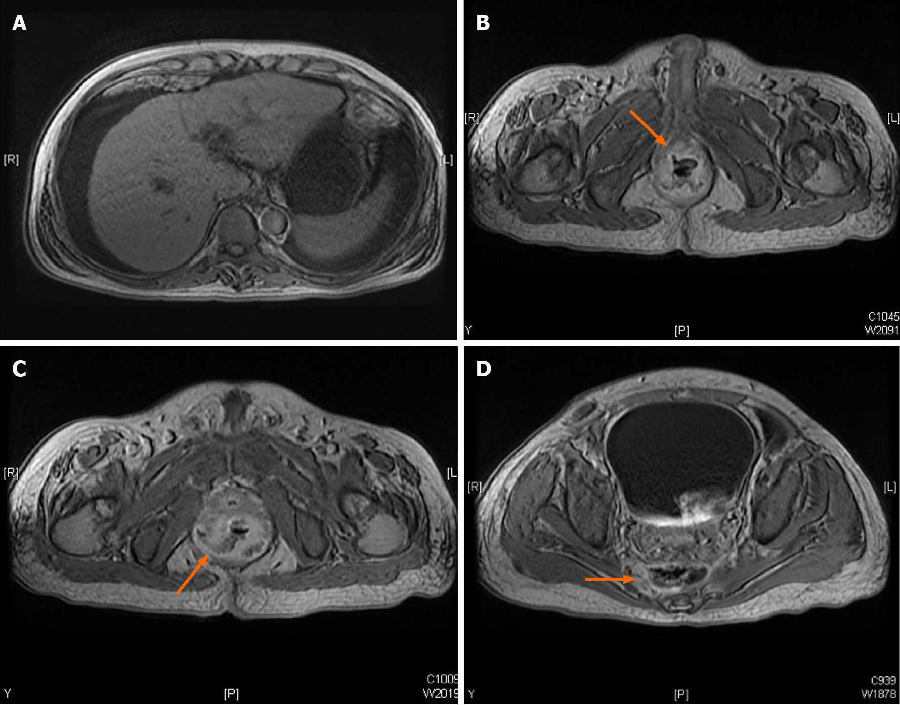
Figure 2 Magnetic resonance imaging at diagnosis.
A: Liver cirrhosis with ascites; B: Rectoprostatic fistula; C: Rectopresacral fistula; D: Presacral abscess.
Figure 3 Pathology.
A: Ulcer with acute on chronic inflammation and granulation tissue at diagnosis; B: Pathological presentations of cytomegalovirus (CMV) infection, immunohistochemistry stain (20 × objective) was performed with 1:200 diluted Novocastra™ lyophilized mouse monoclonal antibody against CMV pp65 antigen and showed strong focal CMV immunoreactivity with brownish areas; C: Minimal inflammatory cells infiltration six months after vedolizumab treatment, seven months after diagnosis.
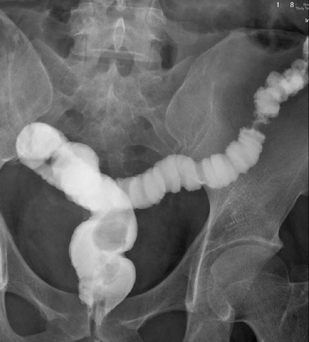
Figure 4 Lower gastrointestinal series showed no more rectal fistula tract.
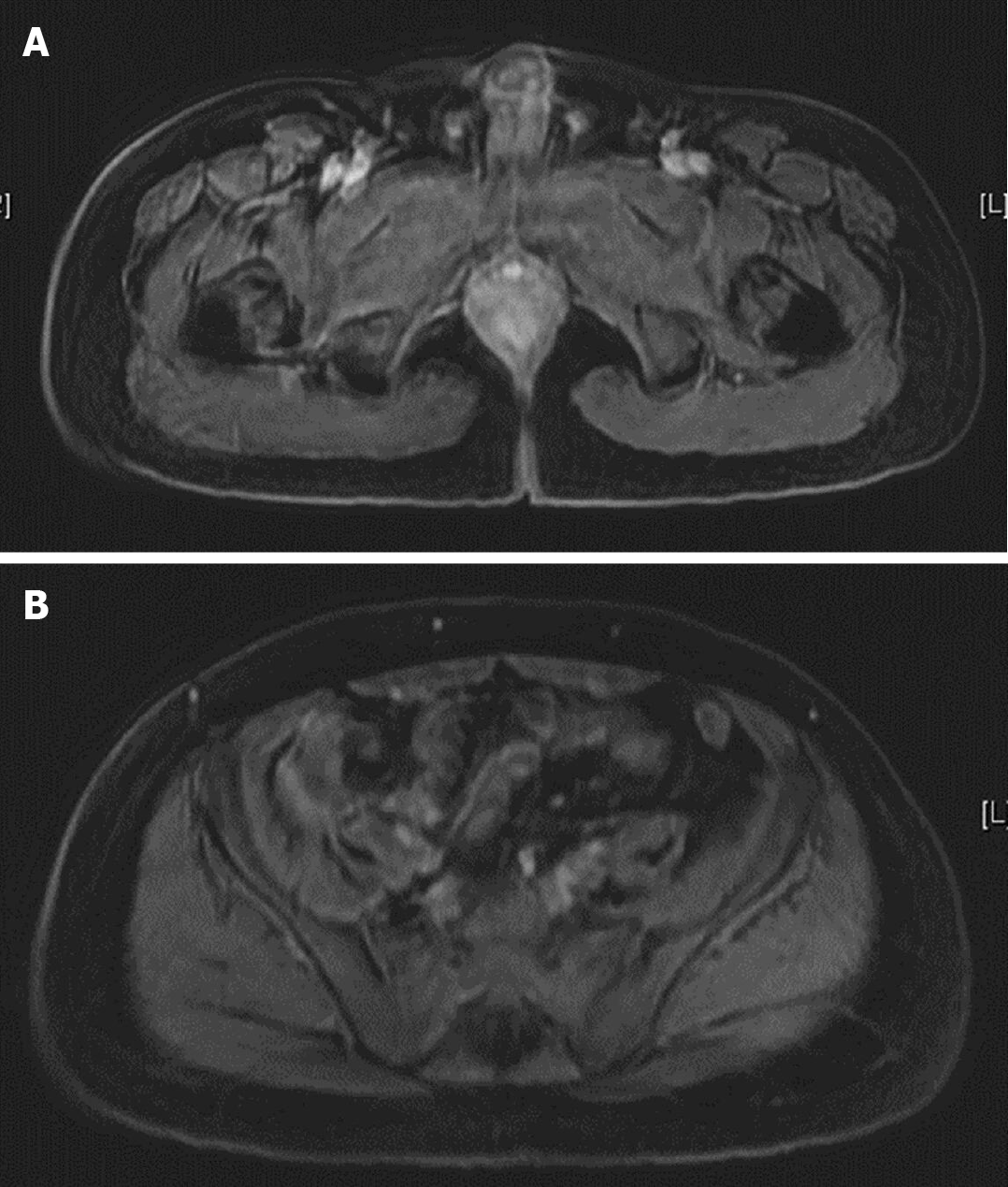
Figure 5 Magnetic resonance imaging seven months after diagnosis.
A: No more rectal fistula tract; B: No more presacral abscess.
- Citation: Yeh H, Kuo CJ, Wu RC, Chen CM, Tsai WS, Su MY, Chiu CT, Le PH. Vedolizumab in Crohn’s disease with rectal fistulas and presacral abscess: A case report. World J Gastroenterol 2021; 27(5): 442-448
- URL: https://www.wjgnet.com/1007-9327/full/v27/i5/442.htm
- DOI: https://dx.doi.org/10.3748/wjg.v27.i5.442


