Copyright
©The Author(s) 2021.
World J Gastroenterol. Dec 28, 2021; 27(48): 8262-8282
Published online Dec 28, 2021. doi: 10.3748/wjg.v27.i48.8262
Published online Dec 28, 2021. doi: 10.3748/wjg.v27.i48.8262
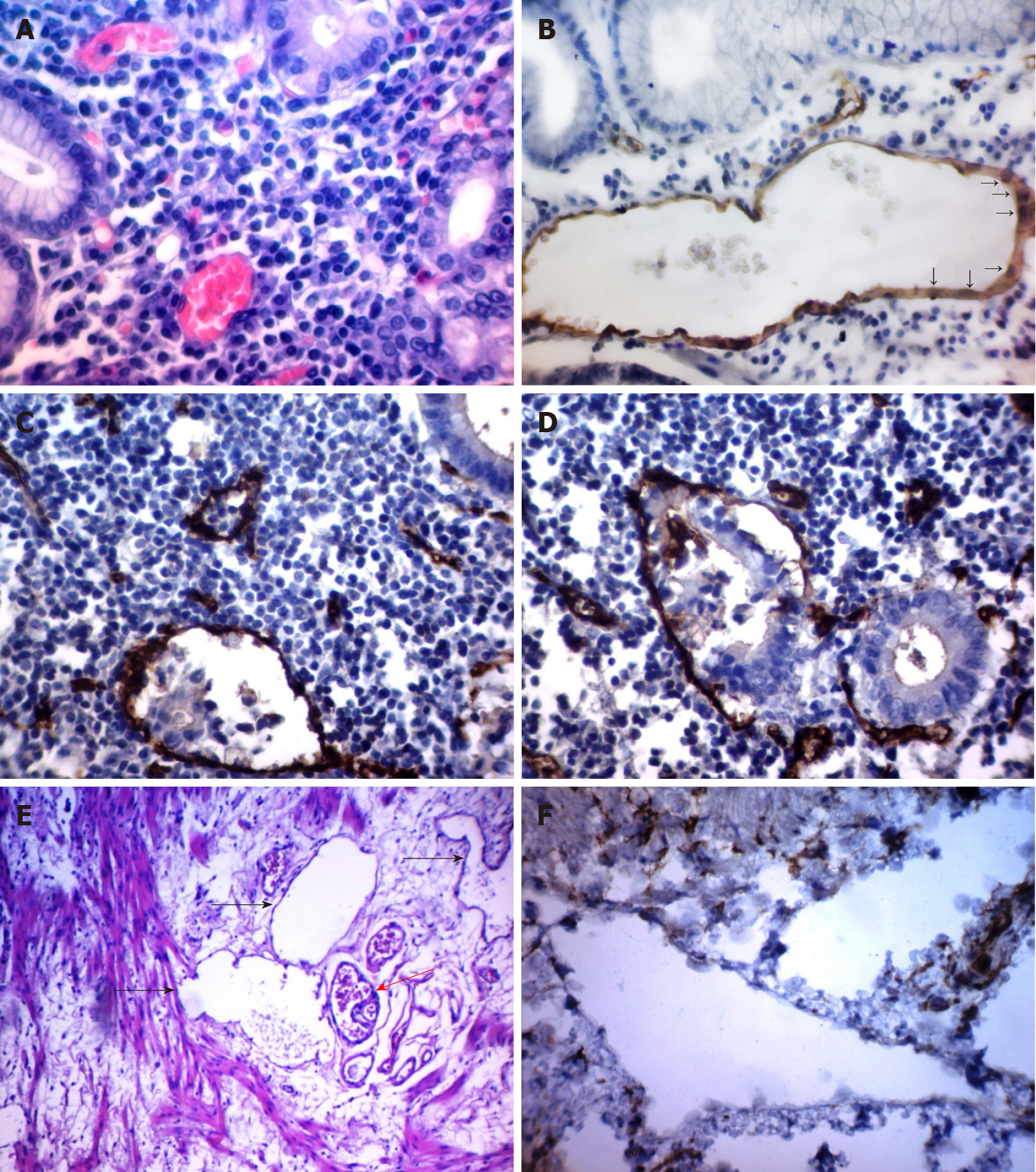
Figure 1 Different types of tumor microvessels in gastric cancer.
A: Normal capillaries in the gastric mucosa adjacent to the tumor [hematoxylin and eosin (HE), 600×]; B: Dilated capillary formed by endothelial cells with large, pale nuclei with fine-netted chromatin structure (arrows) in the gastric mucosa adjacent to the tumor [immunohistochemistry (IHC) staining with antibodies to CD34, 400×]; C: Atypical dilated capillary with tumor emboli in the lumen (IHC staining with antibodies to CD34, 600×); D: Structure with partial endothelial linings (IHC staining with antibodies to CD34, 600×); E: Dilated capillaries with low expression of CD34 (black arrows) and dilated capillary (red arrow) in the gastric submucosa adjacent to the tumor (HE, 200×); F: Dilated capillaries with low expression of CD34 in the gastric submucosa adjacent to the tumor (IHC staining with antibodies to CD34, 600×).
- Citation: Senchukova MA. Issues of origin, morphology and clinical significance of tumor microvessels in gastric cancer. World J Gastroenterol 2021; 27(48): 8262-8282
- URL: https://www.wjgnet.com/1007-9327/full/v27/i48/8262.htm
- DOI: https://dx.doi.org/10.3748/wjg.v27.i48.8262


