Copyright
©The Author(s) 2021.
World J Gastroenterol. Jul 7, 2021; 27(25): 3802-3814
Published online Jul 7, 2021. doi: 10.3748/wjg.v27.i25.3802
Published online Jul 7, 2021. doi: 10.3748/wjg.v27.i25.3802
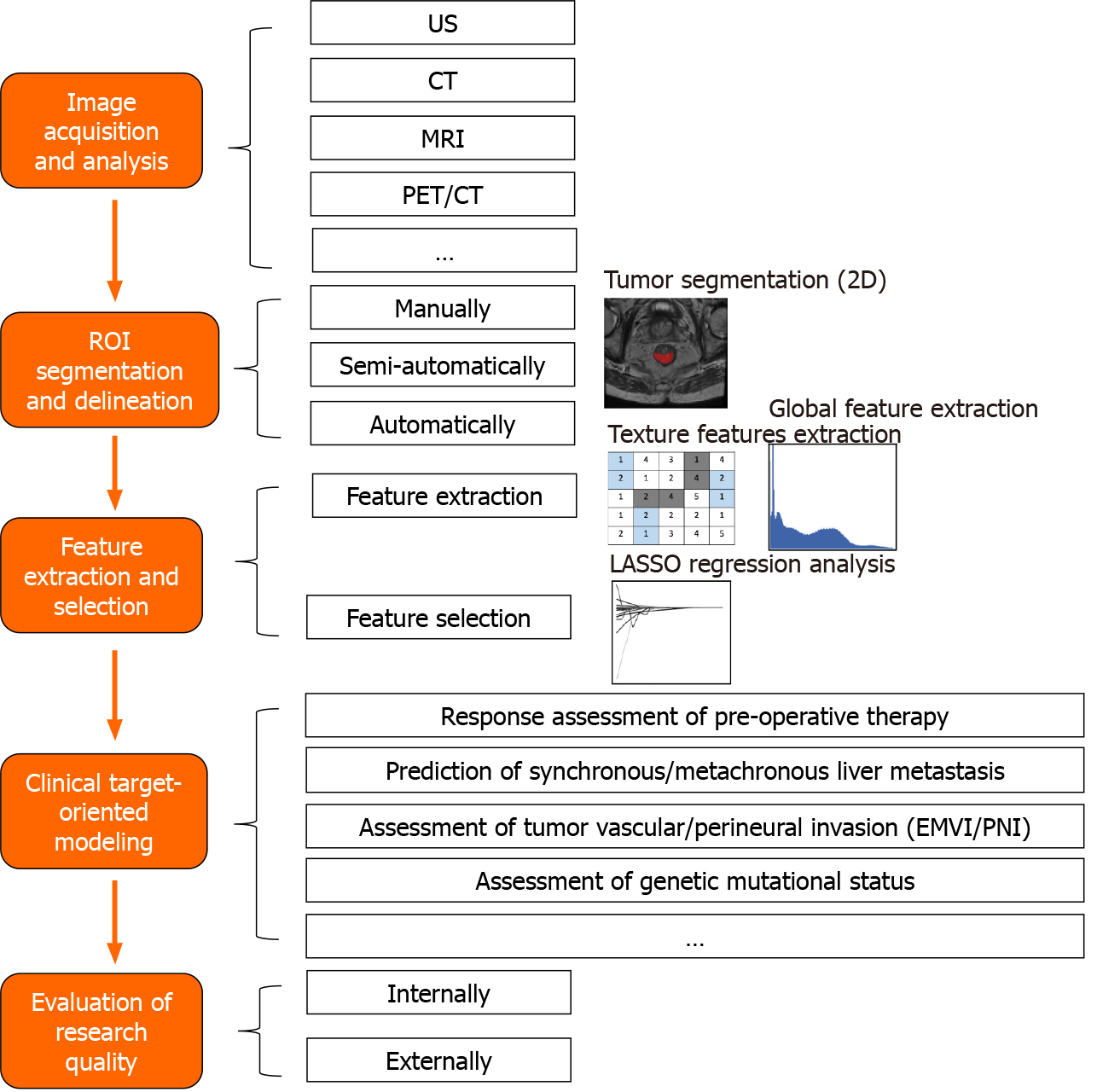
Figure 1 Workflow of radiomics applied in rectal cancer.
US: Ultrasonography; CT: Computed tomography; MRI: Magnetic resonance imaging; PET: Positron emission tomography; ROI: Region of interest; EMVI: Extramural venous invasion; PNI: Perineural invasion.
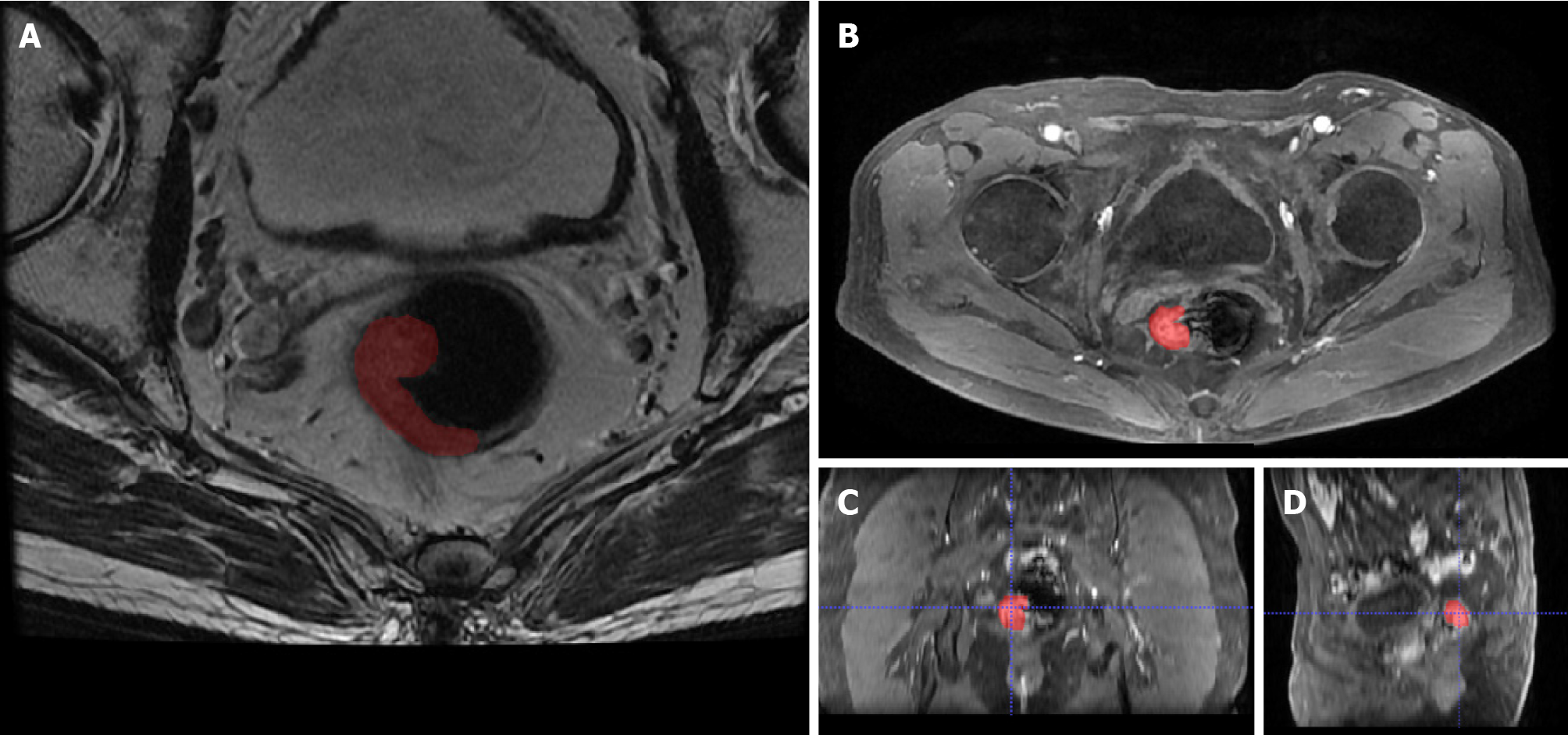
Figure 2 Segmentation of a rectal tumor with Itk-snap software.
A: Example of tumor segmentation using Itk-snap software (www.itksnap.org) on axial plain magnetic resonance image; B-D: Axial (B), reconstructed coronal (C), and sagittal (D) contrast-enhanced magnetic resonance images in the venous phase in a 72-year-old man with rectal cancer.
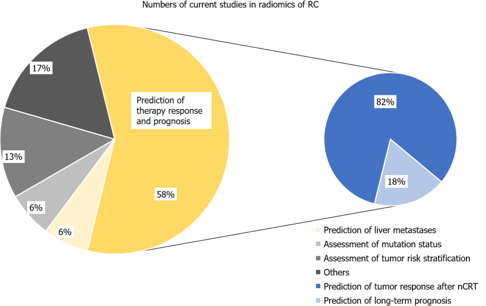
Figure 3 Distribution of the current focus in the industry.
Currently 58% of research focuses on tumor response assessment to preoperative neo-adjuvant radiochemotherapy (nCRT) therapies or prediction of the long-term prognosis, of which most studies are about prediction of tumor response after nCRT. RC: Rectal cancer; nCRT: Neo-adjuvant radiochemotherapy.
- Citation: Hou M, Sun JH. Emerging applications of radiomics in rectal cancer: State of the art and future perspectives. World J Gastroenterol 2021; 27(25): 3802-3814
- URL: https://www.wjgnet.com/1007-9327/full/v27/i25/3802.htm
- DOI: https://dx.doi.org/10.3748/wjg.v27.i25.3802


