Copyright
©The Author(s) 2017.
World J Gastroenterol. Dec 21, 2017; 23(47): 8367-8375
Published online Dec 21, 2017. doi: 10.3748/wjg.v23.i47.8367
Published online Dec 21, 2017. doi: 10.3748/wjg.v23.i47.8367
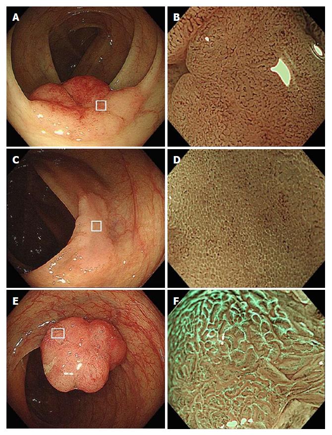
Figure 1 Magnifying endoscopic features with narrow-band imaging.
A: Conventional view. A protruding lesion is detected in the transverse colon. B: M-NBI of the box in Figure 1a shows a clear microvascular pattern without WOS; C: Conventional view. A flat-elevated lesion is detected in the ascending colon. D: M-NBI of box in Figure 1C shows WOS obscuring the microvascular pattern. This WOS is regarded as regular WOS, since it is well-organized and distributed symmetrically with a regular reticular pattern. E: Conventional view. A protruding lesion is detected in the ascending colon. F: M-NBI of the box in Figure 1e shows WOS obscuring the microvascular pattern. This WOS is regarded as irregular WOS, since it is disorganized and distributed asymmetrically with an irregular speckled pattern. M-NBI: Narrow-band imaging; WOS: White opaque substance.
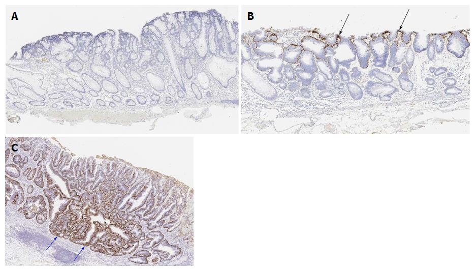
Figure 2 Histopathological findings: Adipophilin immunostaining.
A: Score 0, adipophilin is not detected within the neoplastic epithelium; B: Score 1, adipophilin is detected within the neoplastic epithelium. The depth of adipophilin expression is superficial (black arrows); C: Score 2, adipophilin is detected within the neoplastic epithelium. Adipophilin expression is deep (blue arrows).
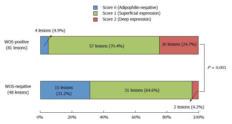
Figure 3 Comparison of the distribution of adipophilin between white opaque substance-positive and -negative lesions.
The scores for adipophilin expression depth are significantly different between the two groups of lesions (P = 0.001), with a predominance of score 2 (deep expression) in WOS-positive lesions (24.7%) compared to WOS-negative lesions (4.2%).
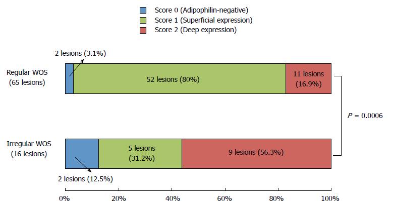
Figure 4 Comparison of the distribution of adipophilin between regular white opaque substance and irregular white opaque substance lesions.
Score 2 (deep expression) is more frequent in lesions with irregular WOS (56.3%) than in lesions with regular WOS (16.9%) (P = 0.0006).
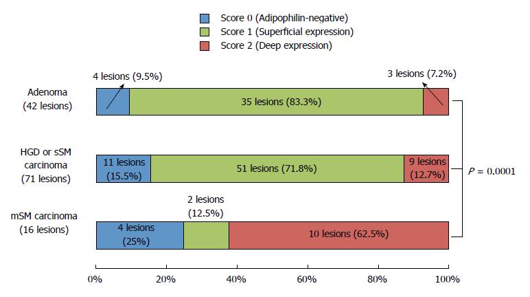
Figure 5 Immunohistochemical findings of colorectal lesions by histologic type.
Score 2 (deep expression) is more frequent in mSM carcinomas (62.5%) than in adenomas (7.2%) or in HGDs/sSM carcinomas (12.7%) (P = 0.0001). HGD: High-grade dysplasia; sSM: Slight submucosal invasion; mSM: Massive submucosal invasion.
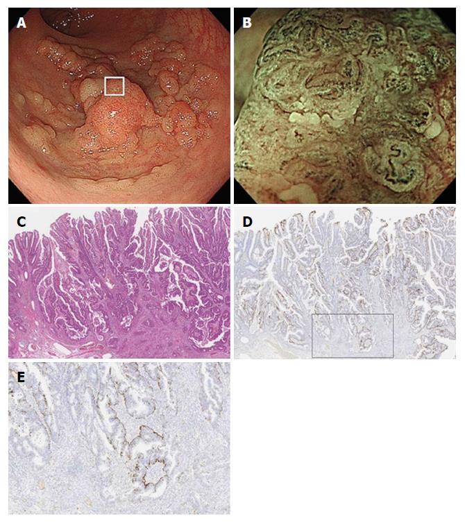
Figure 6 Endoscopic and histologic features of a lesion with irregular white opaque substance.
A: Colonoscopy shows a protruding lesion in the rectum; B: Magnifying endoscopic findings with narrow-band imaging of box in Figure 6a show irregular WOS. WOS is disorganized and asymmetrical; C: Histological examination of the resected specimen shows well to moderately differentiated adenocarcinoma invading the deep submucosal layer (invasion depth; 4,320 μm); D: A low-power view with adipophilin staining shows that the depth of adipophilin expression is deep; E: A high-power view of the box in Figure 6d shows that adipophilin is detected within the neoplastic epithelium. WOS: White opaque substance.
- Citation: Kawasaki K, Eizuka M, Nakamura S, Endo M, Yanai S, Akasaka R, Toya Y, Fujita Y, Uesugi N, Ishida K, Sugai T, Matsumoto T. Association between white opaque substance under magnifying colonoscopy and lipid droplets in colorectal epithelial neoplasms. World J Gastroenterol 2017; 23(47): 8367-8375
- URL: https://www.wjgnet.com/1007-9327/full/v23/i47/8367.htm
- DOI: https://dx.doi.org/10.3748/wjg.v23.i47.8367


