Copyright
©The Author(s) 2017.
World J Gastroenterol. Oct 7, 2017; 23(37): 6911-6919
Published online Oct 7, 2017. doi: 10.3748/wjg.v23.i37.6911
Published online Oct 7, 2017. doi: 10.3748/wjg.v23.i37.6911
Figure 1 Findings from computed tomography.
A: Unenhanced computed tomography (CT) shows a 16 mm × 15 mm, somewhat obscure cystic lesion (red arrow) in the pancreatic tail; B: Contrast-enhanced CT shows an 18 mm × 17 mm cystic lesion (red arrow) with a well-defined, hyper-enhanced, thin peripheral rim and a 6-mm nodule during the arterial phase; C: The outer rim appears iso-enhanced compared to normal pancreatic parenchyma during the portal venous phases (red arrow).
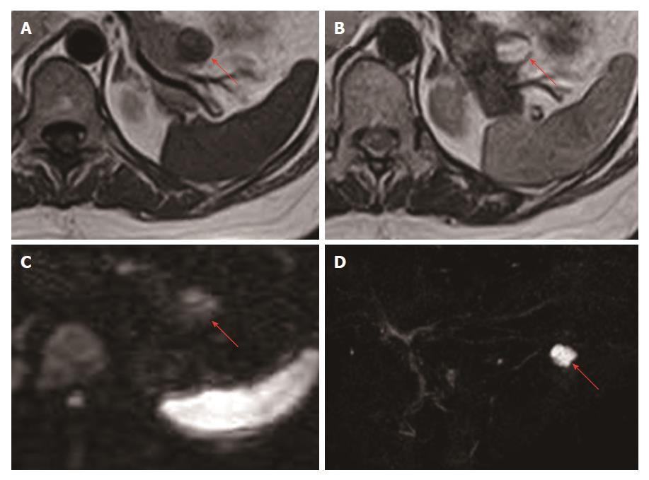
Figure 2 Findings from magnetic resonance imaging.
A: The cystic lesion in the pancreatic tail (red arrow) appears hypointense and some areas appear relatively hyperintense compared to inside the cyst on T1-weighted images; B: The lesion (red arrow) appears mainly hyperintense, while the peripheral rim and nodule appear hypointense on T2-weighted images; C: The lesion (red arrow) appears relatively hyperintense on diffusion-weighted images; D: The lesion (red arrow) is not continuous with the main pancreatic duct on magnetic resonance cholangiopancreatography.
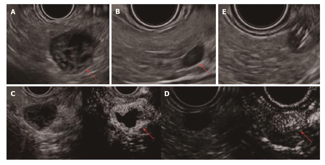
Figure 3 Findings from EUS and CH-EUS.
A: The lesion in the pancreatic tail shows a hypoechoic peripheral rim and nodule. Inside the cyst, no echoic fluid or solid structure is detected; B: A new, 7-mm microtumor in the pancreatic body, apparent only on EUS; C: The rim of the cystic lesion and nodule appear enhanced immediately after injection of contrast medium on CH-EUS; D: The 7-mm microtumor is similarly enhanced on CH-EUS; E: Endoscopic ultrasound-fine needle aspiration to biopsy the 7-mm microtumor. CH-EUS: Contrast-enhanced harmonic-endoscopic ultrasound; EUS: Endoscopic ultrasound.
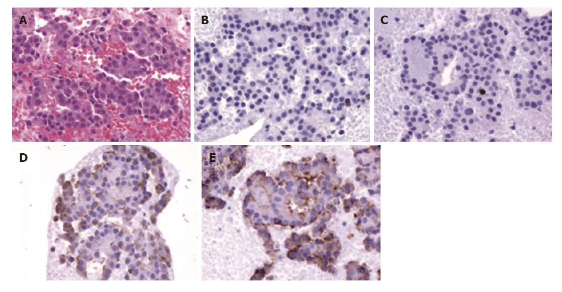
Figure 4 Histopathological findings for the specimen obtained by endoscopic ultrasound-fine needle aspiration.
A: The specimen comprises cells with round nuclei arranged in sheets or rosettes (hematoxylin and eosin, ×100); B and C: Tumor cells show negative results for CD10 (B) and CD56 (C) immunohistochemical staining (×100); D and E: Tumor cells show positive results for chromogranin A (D) and synaptophysin (E) on immunohistochemical staining (×100).
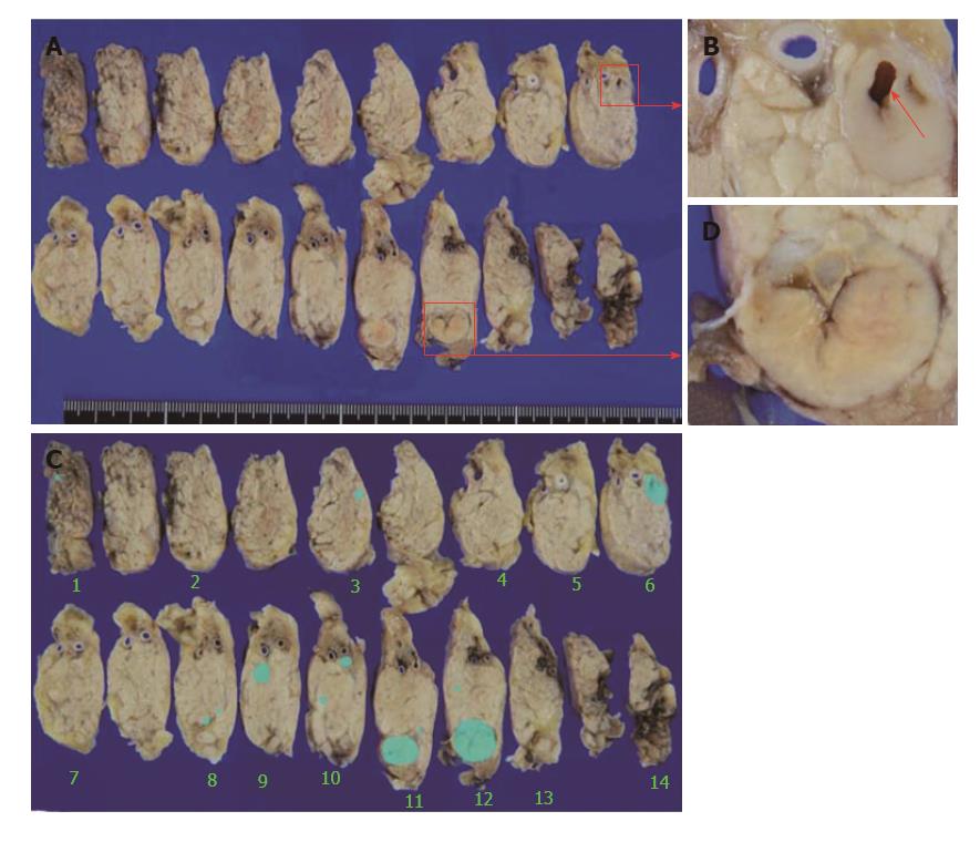
Figure 5 Macroscopic findings for the resected pancreas.
A: The main cystic lesion has morphologically changed to a 13-mm nodule (red square C). The microtumor in the pancreatic body on which EUS-FNA was performed was separate from the main lesion; B: A scar from EUS-FNA is evident in the microtumor (red arrow); C: The main nodule shows internal scarring; D: The main lesion and two microtumors detected on EUS, and another 11 new microtumors (1-3 mm) (blue areas). EUS-FNA: Endoscopic ultrasound-fine needle aspiration.
Figure 6 Microscopic findings for the resected pancreas.
A: The main lesion (red arrow) comprises cells with round nuclei arranged in sheets or rosettes (hematoxylin and eosin, × 4); B: Less than 2 mitoses per 50 high-power fields are evident (hematoxylin and eosin, × 40); C and D: The other microtumors (red arrow) show similar findings (hematoxylin and eosin; C, × 4; D, × 10); E: The peripheral rim appears broken at one site, and fibrosis (red arrow) is evident with inflammation (hematoxylin and eosin, × 10). A trace of a cystic lumen is apparent below the red arrow.
- Citation: Sagami R, Nishikiori H, Ikuyama S, Murakami K. Rupture of small cystic pancreatic neuroendocrine tumor with many microtumors. World J Gastroenterol 2017; 23(37): 6911-6919
- URL: https://www.wjgnet.com/1007-9327/full/v23/i37/6911.htm
- DOI: https://dx.doi.org/10.3748/wjg.v23.i37.6911


