Copyright
©The Author(s) 2017.
World J Gastroenterol. May 28, 2017; 23(20): 3664-3674
Published online May 28, 2017. doi: 10.3748/wjg.v23.i20.3664
Published online May 28, 2017. doi: 10.3748/wjg.v23.i20.3664
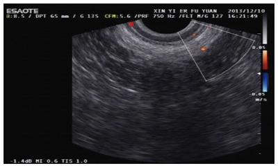
Figure 1 Normal five-layer structure of a healthy participant.
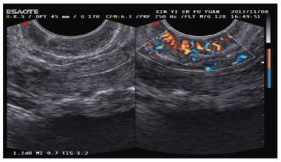
Figure 2 Typical blood flow in patients with stage I or II (left) and stage III or IV (right) hemorrhoids.
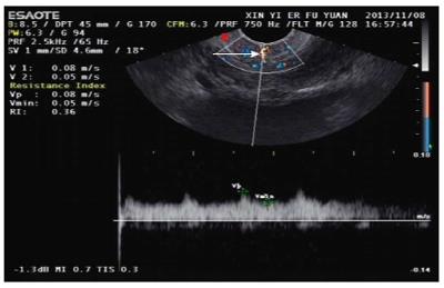
Figure 3 Mosaic pattern in the anal cushion of stages III and IV hemorrhoids.
Blood flow with different directions could be observed as a “mosaic pattern” (white arrow). High-speed low-resistance arterial flow spectrum and arterialized venous spectrum could be observed as a bright colored area.
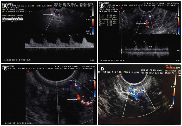
Figure 4 Mosaic pattern in the pathological anal cushion.
A: “Mosaic pattern” was a special blood flow with different directions (white arrow); B: Special blood flow of “mosaic pattern” (white arrow); C: Special blood flow of “mosaic pattern” instructed with red and blue color (white arrow); D: Special blood flow of “mosaic pattern” instructed with multiple colors (white arrow).
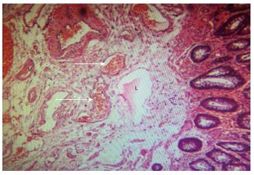
Figure 5 Subepithelial vessels of resected grades III and IV hemorrhoid tissues were manifested by obvious structural impairment and retrograde and ruptured changes of internal elastic lamina.
Arteriovenous fistulas and venous dilatation were obvious in the anal cushion of hemorhoidal tissues (white arrow, magnification × 80); A: Artery, V: Vein, L: Lymphatic duct.
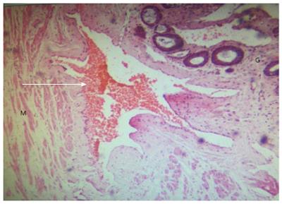
Figure 6 Blood cells in the arteriovenus fistula of anal cushion were seen (white arrow, magnification × 80).
M: Muscle; G: Glands.
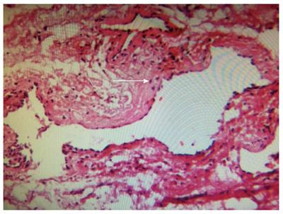
Figure 7 Arteriovenus fistula of anal cushion was obvious (white arrow, magnification × 400).
Some parts of the Trietz’s muscle showed hypertrophy and distortion.
- Citation: Aimaiti A, A Ba Bai Ke Re MMTJ, Ibrahim I, Chen H, Tuerdi M, Mayinuer. Sonographic appearance of anal cushions of hemorrhoids. World J Gastroenterol 2017; 23(20): 3664-3674
- URL: https://www.wjgnet.com/1007-9327/full/v23/i20/3664.htm
- DOI: https://dx.doi.org/10.3748/wjg.v23.i20.3664


