Copyright
©The Author(s) 2017.
World J Gastroenterol. Mar 28, 2017; 23(12): 2246-2250
Published online Mar 28, 2017. doi: 10.3748/wjg.v23.i12.2246
Published online Mar 28, 2017. doi: 10.3748/wjg.v23.i12.2246
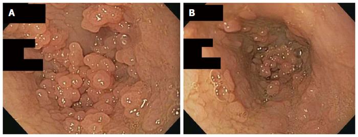
Figure 1 Esophageal endoscopic exam demonstrating multiple papillomas.
A: View of esophageal papillomas in distal esophagus immediately above the level of the lower esophageal sphincter; B: View of the esophageal papillomas and thicken esophageal mucusa in the distal esophagus.
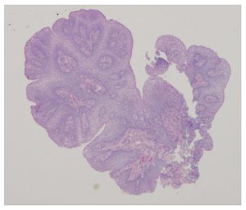
Figure 2 Low power cross section of squamous papilloma obtained from esophagus.
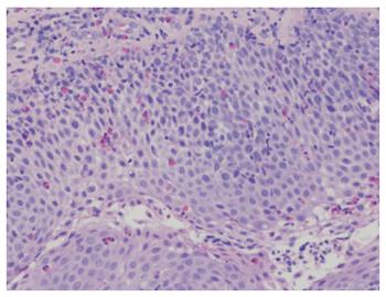
Figure 3 High power view of papilloma demonstrating eosinophilic infiltration.
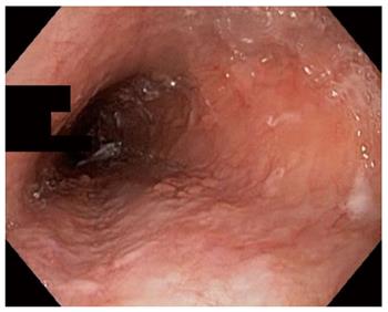
Figure 4 Esophageal endoscopic follow up exam post-treatment with argon plasma coagulation and debulking of the papillomas.
- Citation: Pasman EA, Heifert TA, Nylund CM. Esophageal squamous papillomas with focal dermal hypoplasia and eosinophilic esophagitis. World J Gastroenterol 2017; 23(12): 2246-2250
- URL: https://www.wjgnet.com/1007-9327/full/v23/i12/2246.htm
- DOI: https://dx.doi.org/10.3748/wjg.v23.i12.2246


