Copyright
©The Author(s) 2016.
World J Gastroenterol. Mar 7, 2016; 22(9): 2760-2770
Published online Mar 7, 2016. doi: 10.3748/wjg.v22.i9.2760
Published online Mar 7, 2016. doi: 10.3748/wjg.v22.i9.2760
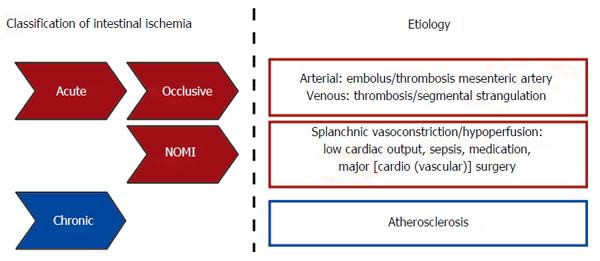
Figure 1 Schematic overview of intestinal ischemia based on etiological background.
NOMI: Nonocclusive mesenteric ischemia.
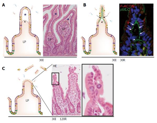
Figure 2 The human small intestine has efficient mechanisms to prevent excessive epithelial lining damage.
A: Cartoon and hematoxylin-eosin staining demonstrating the appearance of subepithelial spaces (asterisks) after ischemia as a result of retraction of the basement membrane (arrowhead); B: Early during reperfusion, loose IR-damaged epithelial sheets are pulled together through active contraction of pMLC at the basal side of epithelial cells (green line, arrows), bringing these cells together; C: Zipper-like constriction of the epithelium is associated with rapid restoration of the epithelial lining and prevents exposure of lamina propria to intraluminal content. LP: Lamina propria; pMLC: Phosphorylated myosin light chain; F-ACTIN: Filamentous actin.
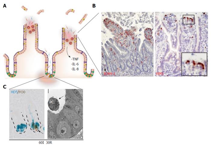
Figure 3 Prolonged small intestinal ischemia-reperfusion results in physical and immunological barrier function loss and inflammation.
A: Prolonged IR leads to disruption of the epithelial lining (physical barrier integrity loss) and Paneth cell loss (immunological barrier integrity loss); B: Inflammatory responses are characterized by increased endothelial expression of ICAM-1 (left panel) with sequestration of MPO-positive neutrophils into the villus tips (right panel), and increased expression and release of inflammatory cytokines including IL-6, IL-8 and TNF; C: Left panel: Prolonged IR leads to Paneth cell apoptosis, as shown by the co-localization of M30 (brown: apoptosis) and human defensin 5 (blue: Paneth cells). Right panel: EM picture of apoptotic Paneth cell, shed into the crypt lumen. ICAM-1: Intercellular adhesion molecule-1; MPO: Myeloperoxidase; HD5: Human defensin-5.
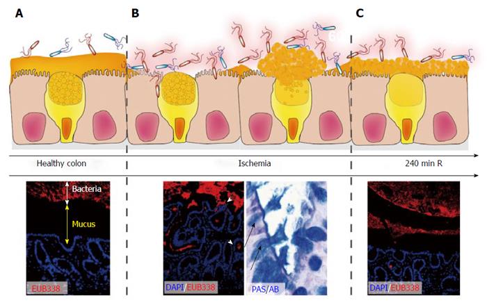
Figure 4 Rapid restoration of colon ischemia-reperfusion-induced mucus barrier loss by goblet cell compound exocytosis.
In healthy colon, bacteria are separated from the epithelium by a thick mucus layer (A; DAPI, Blue: nuclei. EUB338, Red: bacteria); During ischemia and early reperfusion, the mucus barrier is damaged leading to penetration of bacteria deep into the colonic crypts (B). Goblet cells respond by massive release of their granules into the crypt lumen (so called compound exocytosis), which led to a recoverd mucus layer at 240 min of reperfusion in the rat intestine (C). Note that bacterial clearance is accompanied by depletion of goblet cell contents.
- Citation: Grootjans J, Lenaerts K, Buurman WA, Dejong CHC, Derikx JPM. Life and death at the mucosal-luminal interface: New perspectives on human intestinal ischemia-reperfusion. World J Gastroenterol 2016; 22(9): 2760-2770
- URL: https://www.wjgnet.com/1007-9327/full/v22/i9/2760.htm
- DOI: https://dx.doi.org/10.3748/wjg.v22.i9.2760


