Copyright
©The Author(s) 2016.
World J Gastroenterol. Mar 21, 2016; 22(11): 3296-3301
Published online Mar 21, 2016. doi: 10.3748/wjg.v22.i11.3296
Published online Mar 21, 2016. doi: 10.3748/wjg.v22.i11.3296
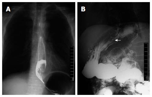
Figure 1 Upper gastrointestinal barium examination showed a large tumor blocking the esophago-gastric junction (A), esophagram showed a niche shadow in gastric fundus (B).
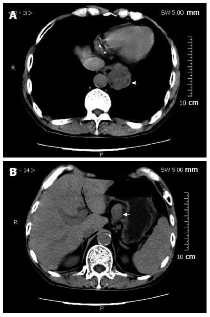
Figure 2 Computed tomography scan showed a soft mass in the esophago-gastric junction (A), enlarged lymph nodes in the lesser curvature of the stomach (B).
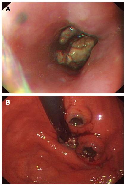
Figure 3 Endoscopic examination showed a black spot in the lower esophagus and a bulky black mass blocking the esophago-gastric junction (A), two black crater-like ulcers located at the fundus of the stomach (B).
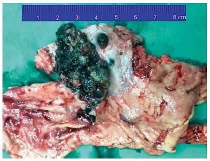
Figure 4 Surgical specimen showed that the melanoma measured 3 cm × 6 cm in size with black pigmentation.
There were several pigmented satellite nodules beside the main tumor lesion, the largest one being 1 cm × 1 cm in diameter. Two ulceration lesions were present at the fundus of the stomach.
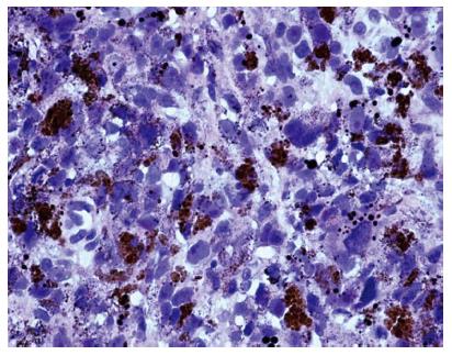
Figure 5 Histological examination showed that the excised tumor tissue was composed of non-organized and pleomorphic cells exhibiting atypical nuclei, and abundant melanin granules.
Hematoxylin-eosin staining, magnification × 400.
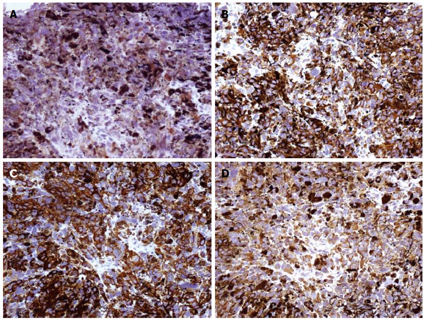
Figure 6 Immunohistochemical staining showed that the tumor was positive for S-100 (A), HMB-45 (B), melan-A (C), and Vimentin (D).
- Citation: Wang L, Zong L, Nakazato H, Wang WY, Li CF, Shi YF, Zhang GC, Tang T. Primary advanced esophago-gastric melanoma: A rare case. World J Gastroenterol 2016; 22(11): 3296-3301
- URL: https://www.wjgnet.com/1007-9327/full/v22/i11/3296.htm
- DOI: https://dx.doi.org/10.3748/wjg.v22.i11.3296


