Copyright
©2014 Baishideng Publishing Group Inc.
World J Gastroenterol. Jun 28, 2014; 20(24): 7794-7800
Published online Jun 28, 2014. doi: 10.3748/wjg.v20.i24.7794
Published online Jun 28, 2014. doi: 10.3748/wjg.v20.i24.7794
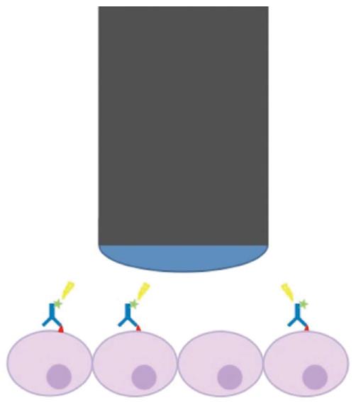
Figure 1 Molecular imaging with labelled antibodies using the confocal laser endomicroscopy system.
Antibodies (blue) labelled with a fluorochrome (FITC or Alexa-Fluor 488) (green) bind to a specific target (red triangles) expressed on the plasma membrane of cells in the mucosal layer in the gastrointestinal tract (purple). The confocal laser endomicroscopy (CLE) scope (black and blue tube) excites the fluorochrome with a defined wavelength of 488 nm (yellow). Light is subsequently emitted and detected by the CLE scope.
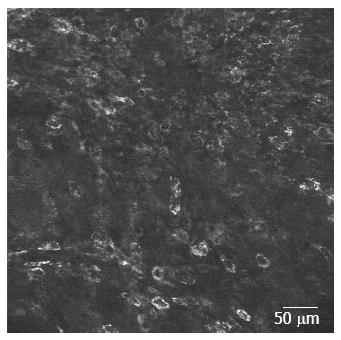
Figure 2 In vivo epidermal growth factor receptor staining in a human colorectal cancer xenograft (SW480) after intratumoural injection of fluorescein isothiocyanate-labelled anti-epidermal growth factor receptor-antibody.
The edge length is 475 μm. Courtesy of Dr. M. Hoetker and Prof. M. Goetz, Tuebingen, Germany.
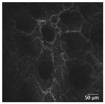
Figure 3 Molecular confocal laser endomicroscopy staining with Alexa-Fluor 488 labelled anti-CD31 antibodies.
As CD31 is an endothelial marker, the structures seen in the image are microvessels in a colonic adenocarcinoma.
-
Citation: Karstensen JG, Klausen PH, Saftoiu A, Vilmann P. Molecular confocal laser endomicroscopy: A novel technique for
in vivo cellular characterization of gastrointestinal lesions. World J Gastroenterol 2014; 20(24): 7794-7800 - URL: https://www.wjgnet.com/1007-9327/full/v20/i24/7794.htm
- DOI: https://dx.doi.org/10.3748/wjg.v20.i24.7794


