Copyright
©The Author(s) 1996.
World J Gastroenterol. Jun 25, 1996; 2(2): 82-85
Published online Jun 25, 1996. doi: 10.3748/wjg.v2.i2.82
Published online Jun 25, 1996. doi: 10.3748/wjg.v2.i2.82
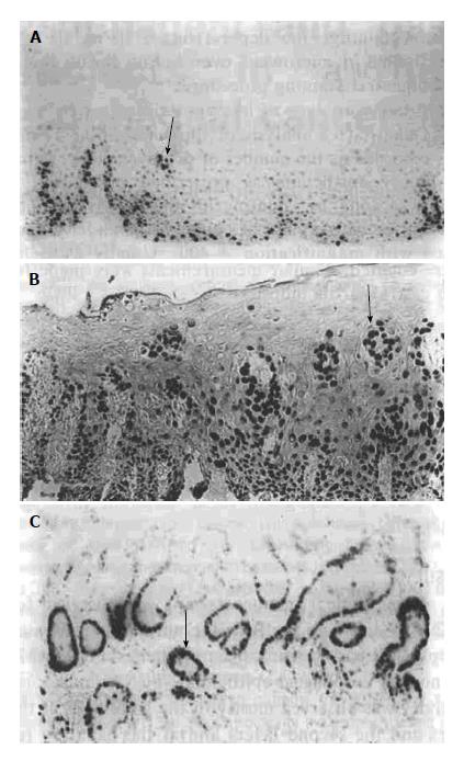
Figure 1 Immunostaining of Proliferating cell nuclear antigen in biopsied samples of esophageal and gastric cardia epithelia.
Immunoreactivity is located in the nuclei of basal cells in the papillary region of the normal epithelia of esophagus (A, arrows) and the positive cells expand in the upper region of DYS (B). C: PCNA staining in gastric cardia epithelium with CSG (× 100). PCNA: Proliferating cell nuclear antigen; DYS: Dysplasia; CSG: Chronic superficial gastritis.
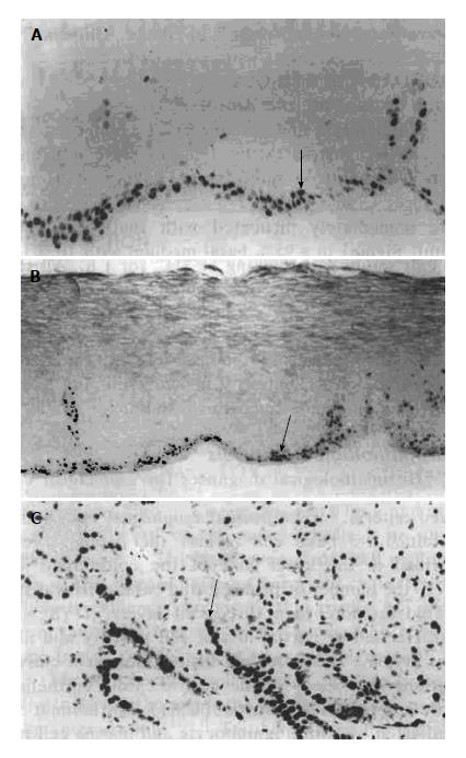
Figure 2 Immunostaining of Ki-67 in biopsied samples of esophageal and gastric cardia epithelia.
Immunoreactivity is located in the nuclei of basal cells in the papillary region of the normal epithelia of esophagus (A, arrows) and the positive cells expand in the upper region of DYS (B). C: Ki-67 staining in gastric cardia epithelium with CSG (× 100). Immunoreactivity is located in the nuclei of cells at the basal cells of the crypts and at the deep glands (arrows). DYS: Dysplasia.
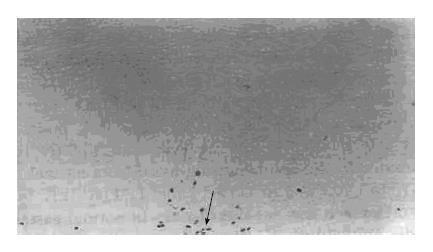
Figure 3 Immunohistochemical studies of BudR-labeled cells in the esophageal epithelia of basal cell hyperplasia (with methyl green counterstaining).
Immunoreactivity is located in the cell nuclei (× 200, arrows).
- Citation: Wang LD, Zhou Q, Gao SS, Li YX, Yang WC. Measurements of cell proliferation in esophageal and gastric cardia epithelia of subjects in a high incidence area for esophageal cancer. World J Gastroenterol 1996; 2(2): 82-85
- URL: https://www.wjgnet.com/1007-9327/full/v2/i2/82.htm
- DOI: https://dx.doi.org/10.3748/wjg.v2.i2.82


