Copyright
©2013 Baishideng Publishing Group Co.
World J Gastroenterol. Oct 21, 2013; 19(39): 6699-6702
Published online Oct 21, 2013. doi: 10.3748/wjg.v19.i39.6699
Published online Oct 21, 2013. doi: 10.3748/wjg.v19.i39.6699
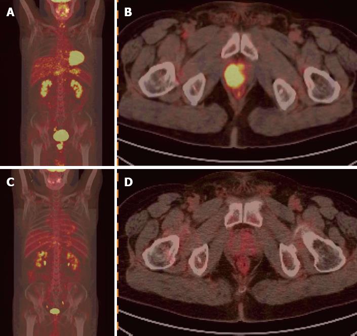
Figure 1 Positron emission tomography/computerized tomography image.
A: Whole body positron emission tomography (PET) image (scan) shows nodal hypermetabolic foci in the prostate, and no abnormally high uptake foci in the other sites of the body; B: The PET/computerized tomography (CT) fusion image displays a nodal hypermetabolic lesion located in the right lobe of the prostate, with maximal standardized uptake value (SUV) being 12.7; C: There were no hypermetabolic foci on the whole body PET imaging following six courses of chemotherapy; D: There was complete remission of the prostatic lesion after six cycles of chemotherapy, and the maximal SUV was 2.0.
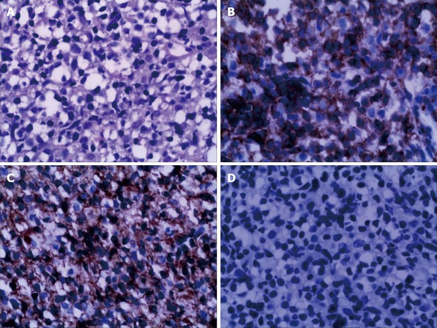
Figure 2 Combined morphological and immunophenotyping examinations confirmed a diffuse large B cell lymphoma of non-germinal center B-cell origin.
A: Intensive proliferation of lymphoid cells (Hematoxylin and eosin, × 400); B and C: Diffuse and consistent expression of CD79a, CD20 (immunohistochemistry, × 400); D: The expression of serum prostate-pecific antigen is negative (immunohistochemistry, × 400).
- Citation: Pan B, Han JK, Wang SC, Xu A. Positron emission tomography/computerized tomography in the evaluation of primary non-Hodgkin’s lymphoma of prostate. World J Gastroenterol 2013; 19(39): 6699-6702
- URL: https://www.wjgnet.com/1007-9327/full/v19/i39/6699.htm
- DOI: https://dx.doi.org/10.3748/wjg.v19.i39.6699


