Copyright
©2013 Baishideng Publishing Group Co.
World J Gastroenterol. Sep 28, 2013; 19(36): 6000-6010
Published online Sep 28, 2013. doi: 10.3748/wjg.v19.i36.6000
Published online Sep 28, 2013. doi: 10.3748/wjg.v19.i36.6000
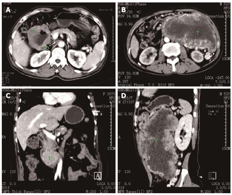
Figure 1 Respective computed tomography images.
A: For case 28; B, D: For case 20; C: For case 18. (T: Tumor; D: Duodenum).
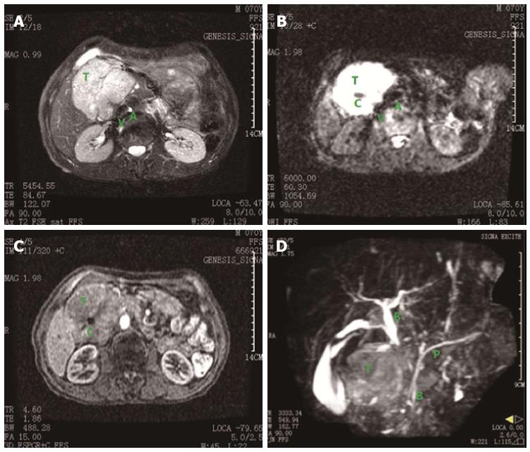
Figure 2 Respective magnetic resonance imaging images.
A-C: For case 4; D: For case 22. T: Tumor; C: Necrotic core; A: Abdominal aorta; V: Inferior vena cava; B: Common bile duct; P: Main pancreatic duct.
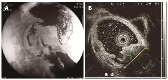
Figure 3 Respective gastrointestinal and endoscopic ultrasonography images.
A: Gastrointestinal for case 4; B: Endoscopic ultrasonography for case 23. T: Tumor.
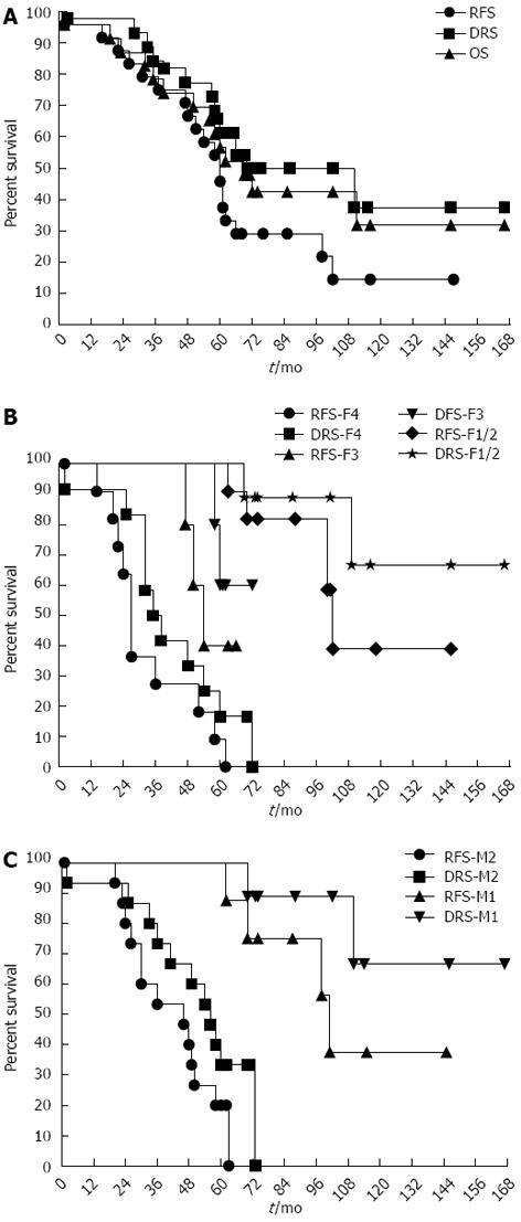
Figure 4 Survival curves.
DRS: Disease-related survival; RFS: Relapse-free survival; OR: Overall survival; DFS: Disease free survival.
- Citation: Liang X, Yu H, Zhu LH, Wang XF, Cai XJ. Gastrointestinal stromal tumors of the duodenum: Surgical management and survival results. World J Gastroenterol 2013; 19(36): 6000-6010
- URL: https://www.wjgnet.com/1007-9327/full/v19/i36/6000.htm
- DOI: https://dx.doi.org/10.3748/wjg.v19.i36.6000


