Copyright
©2010 Baishideng.
World J Gastroenterol. May 21, 2010; 16(19): 2355-2361
Published online May 21, 2010. doi: 10.3748/wjg.v16.i19.2355
Published online May 21, 2010. doi: 10.3748/wjg.v16.i19.2355
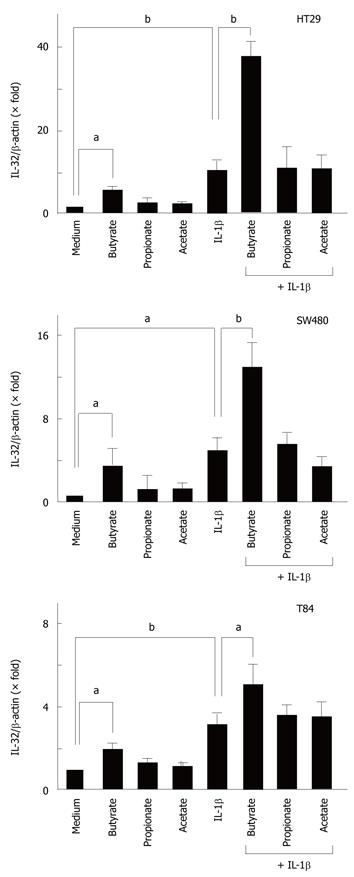
Figure 1 Effects of short-chain fatty acids (SCFAs) on IL-32α mRNA expression in colon cancer cell lines.
The cell lines (HT-29, SW480, T84) were incubated for 12 h with each SCFA (10 mmol/L). In another experiment, the cells were stimulated with IL-1β (50 ng/mL) in the presence or absence of each SCFA (10 mmol/L). The IL-32α mRNA expression was determined by real-time PCR. The data are expressed as mean ± SD (n = 4). aP < 0.05, bP < 0.01.
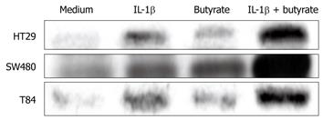
Figure 2 Effects of SCFAs on IL-32α protein secretion in colon cancer cell lines.
The cell lines (HT-29, SW480, T84) were stimulated for 48 h with IL-1β (50 ng/mL) in the presence or absence of butyrate (10 mmol/L), and the intracellular IL-32α protein levels were detected by Western blotting.
Figure 3 Dose-dependent effects of butyrate on IL-1β-induced IL-32α mRNA expression in colon cancer cell lines.
The cell lines (HT-29, SW480, T84) were stimulated for 12 h with increasing concentrations (0-10 mmol/L) of butyrate in the presence or absence of IL-1β (50 ng/mL). IL-32α mRNA expression was then analyzed by real-time PCR. The data are expressed as mean ± SD (n = 4). aP < 0.05, cP < 0.005.
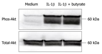
Figure 4 Effects of butyrate on Akt phosphorylation.
The cells were stimulated in the presence of butyrate (5 mmol/L) and/or IL-1β (50 ng/mL) for 15 min, and then Akt phosphorylation was determined by Western blotting.
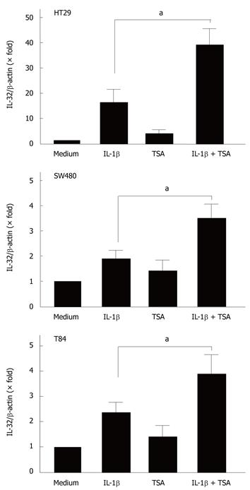
Figure 5 Effects of trichostatin-A (TSA) on IL-32α mRNA expression.
The cells (HT-29, SW480, T84) were incubated for 12 h with IL-1β (50 ng/mL) in the presence or absence of TSA (5 μmol/L), and then the IL-32α mRNA expression was determined by real-time PCR. The data are expressed as mean ± SD (n = 4). aP < 0.05.
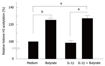
Figure 6 Effects of butyrate on histone H3 acetylation.
HT-29 cells were stimulated for 12 h with IL-1β (50 ng/mL) in the presence or absence of butyrate (10 mmol/L). Histone H3 acetylation was then detected by histone H3 acetylation assay kits (Epigentek; Brooklyn, NY, USA). Histone H3 acetylation was expressed as a value relative to medium alone. The data are expressed as mean ± SD (n = 4). aP < 0.05, bP < 0.01.
- Citation: Kobori A, Bamba S, Imaeda H, Ban H, Tsujikawa T, Saito Y, Fujiyama Y, Andoh A. Butyrate stimulates IL-32α expression in human intestinal epithelial cell lines. World J Gastroenterol 2010; 16(19): 2355-2361
- URL: https://www.wjgnet.com/1007-9327/full/v16/i19/2355.htm
- DOI: https://dx.doi.org/10.3748/wjg.v16.i19.2355


