Copyright
©2010 Baishideng.
World J Gastroenterol. May 14, 2010; 16(18): 2302-2304
Published online May 14, 2010. doi: 10.3748/wjg.v16.i18.2302
Published online May 14, 2010. doi: 10.3748/wjg.v16.i18.2302
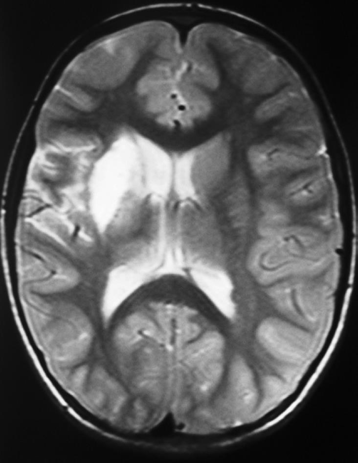
Figure 1 Brain magnetic resonance imaging shows an infarction area measuring 31 mm × 14 mm at the right basal ganglia level.
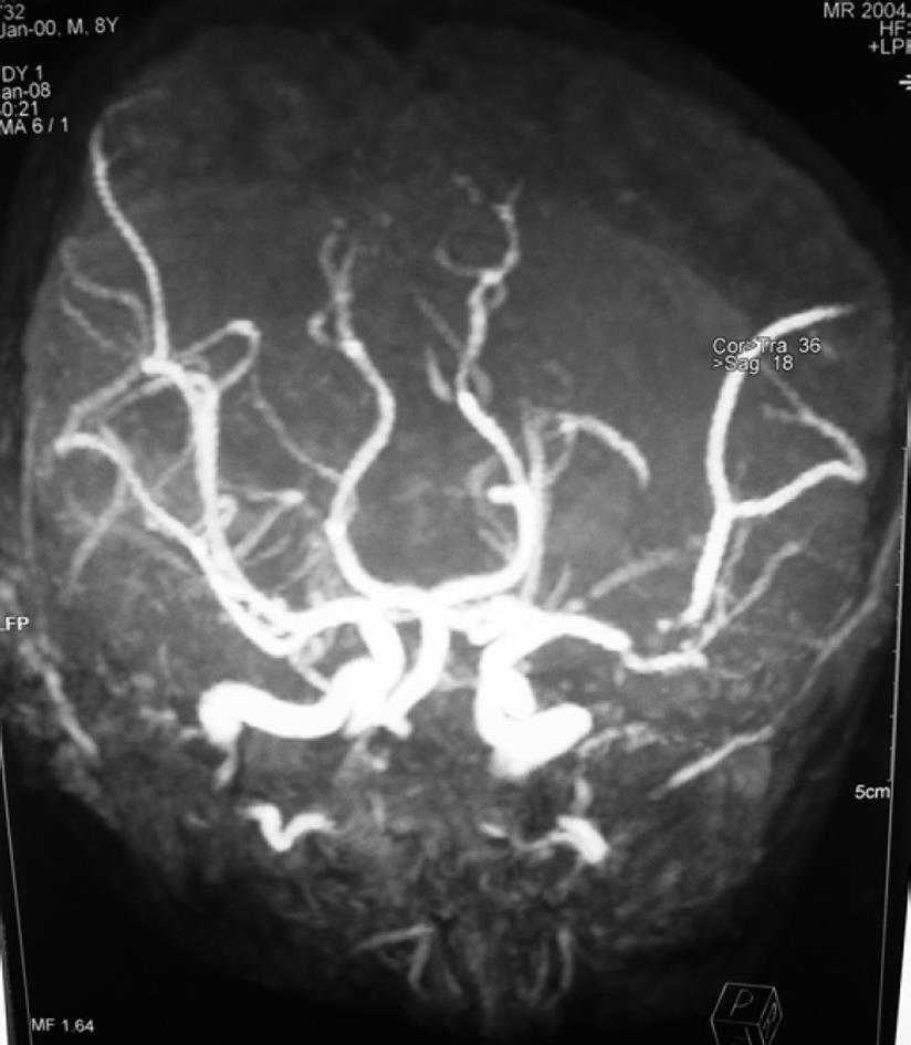
Figure 2 Cerebral angiography examination shows a 1 cm segment occlusion at the M2 branch of the right middle cerebral artery.
- Citation: Doğan M, Peker E, Cagan E, Akbayram S, Acikgoz M, Caksen H, Uner A, Cesur Y. Stroke and dilated cardiomyopathy associated with celiac disease. World J Gastroenterol 2010; 16(18): 2302-2304
- URL: https://www.wjgnet.com/1007-9327/full/v16/i18/2302.htm
- DOI: https://dx.doi.org/10.3748/wjg.v16.i18.2302


