Copyright
©The Author(s) 2017.
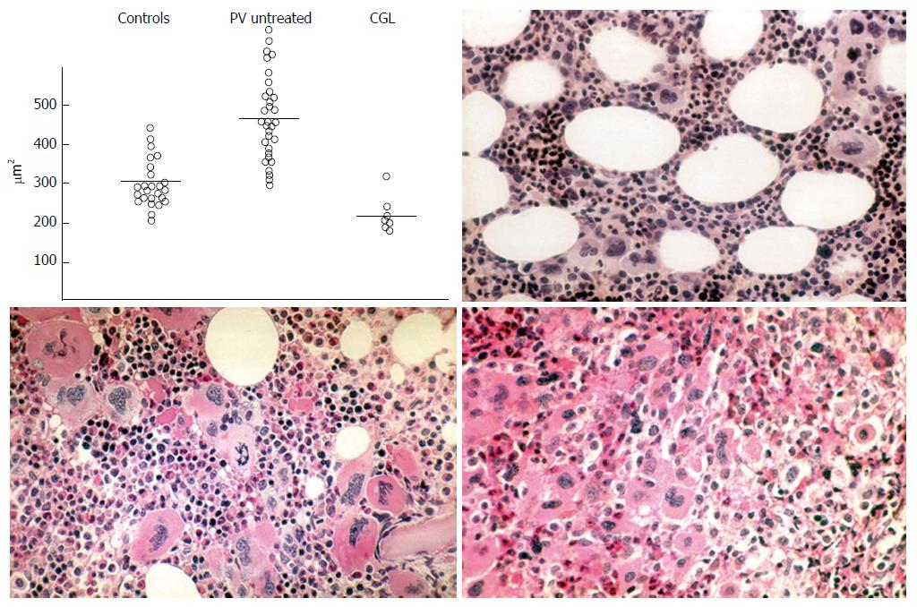
Figure 5 Planimetry of megakaryocyte sizes (μm2) from bone marrow smears in controls, polycythemia vera and chronic granulocytic leukemia upper left: Normal size megakaryocytes in controls; large megakaryocytes in untreated polycythemia vera and small sized megakaryocytes in chronic granulocytic leukemia (Frantzen et al[40]).
Demonstration by Michiels (1981) of a spectrum of clustered large megakaryocytes with hyperlobulated nuclei and a normocellular bone marrow in essential thrombocythemia (ET) vs increased bone marrow cellularity duet o increased erythropoiesis in ET and polycythemia vera (PV) vs increased trilinear eythrocythemic, megakaryocythemia and granulocythemic (EMG) proliferation in classical PV according to Dameshek[38] (1950) and Kurnicke et al[39].
- Citation: Michiels JJ. Aspirin cures erythromelalgia and cerebrovascular disturbances in JAK2-thrombocythemia through platelet-cycloxygenase inhibition. World J Hematol 2017; 6(3): 32-54
- URL: https://www.wjgnet.com/2218-6204/full/v6/i3/32.htm
- DOI: https://dx.doi.org/10.5315/wjh.v6.i3.32


