Copyright
©The Author(s) 2016.
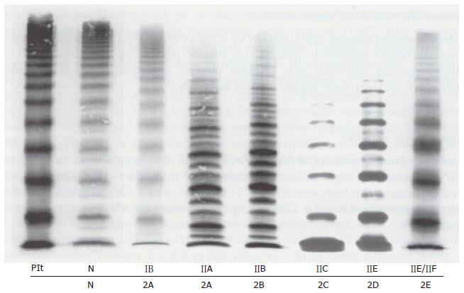
Figure 6 Multimeric pattern.
VWF from plasma of patients with von Willebrand disease classified according to the ISTH criteria for VWD type IIA, IIB, IIC and IIE and the translation into the 2001 Hamburg criteria for 1B = 2M (not 2A), 2A, 2B, 2C, 2D and 2E anno 2001 compared to a normal control in high resolution gel concentration (1.5%) according to Schneppenheim et al[10]. Dominant 1B relatively lacking large VWF multimers (MM) = 2M (Michiels). Dominant IIA = 2A lack of large molecular weight MM and the outer sub-bands of the individual triplets are markedly pronounced indicating increased proteolysis as the cause of 2A. Dominant 2B cannot be distinguished from 2A by MM alone. Recessive IIC = 2C lack of large MM and absence of triplets. Low MM and especially the first band, which probably reflects protomer (dimer) and a tetramer, is markedly pronounced. IID = 2D intervening VWF band and an odd number of MM. 2E lack or relative decrease of large MM and absence of the outer sub-bands of the normal triplet structure. Triplets are lacking in 2C, 2D, 2E and 1B = 2M are lacking indicating the absence of proteolysis. VWD: Von Willebrand disease; VWF: Von Willebrand factor. Plt: Platelet VWF; N: Normal.
- Citation: Michiels JJ, Batorova A, Prigancova T, Smejkal P, Penka M, Vangenechten I, Gadisseur A. Changing insights in the diagnosis and classification of autosomal recessive and dominant von Willebrand diseases 1980-2015. World J Hematol 2016; 5(3): 61-74
- URL: https://www.wjgnet.com/2218-6204/full/v5/i3/61.htm
- DOI: https://dx.doi.org/10.5315/wjh.v5.i3.61


