Copyright
©The Author(s) 2016.
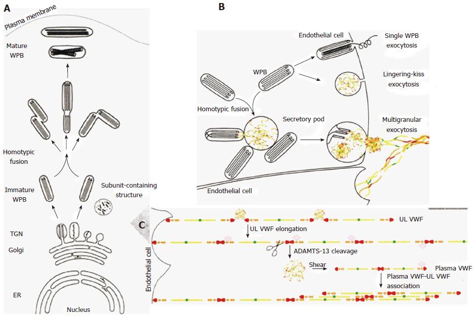
Figure 3 A left biosynthesis pathway of Weibel-Palade body[4].
A, B: The different steps in WPB synthesis of von Willebrand factor (VWF) assembly at the level of endoplasmatic reticulum (ER), at the trans-Golgi network (TGN) level (A), and VWF tubules are assembled and packed into budding vesicles prior to immature WPB formation. Homotypic fusion of WPB gives rise to the formation of WPB with different shapes. As WPB mature they became more electron-dense and reach the plasma membrane. B: Different modes of WPB exocytosis and VWF string formation on endothelial cells. In single WPB exocytosis mode, a single WPB fuses with the plasma membrane and ultra large VWF multimers (MM) are secreted. In lingering-kiss exocytosis mode (B), WPB round up and a small pore is formed with the plasma membrane, allowing the secretion of ultra large VWF MM. In multigranular exocytosis mode (B), WPB undergo homotypic fusion leading to the formation of a secretory pod that permits pooling of ultra large VWF MM prior to secretion[4]. After release, the ultra large vWF strings stick to the endothelial cell surface, attract platelets through platelet GpIb ligand and VWF GpIb receptor interaction, thereby activating the VWF cleavage site to be cleaved by ADAMTS13 at high shear stress in the endarterial circulation (C). WPB: Weibel-palade body. Source: Valentijn and Eikenboom 2013[4].
- Citation: Michiels JJ, Batorova A, Prigancova T, Smejkal P, Penka M, Vangenechten I, Gadisseur A. Changing insights in the diagnosis and classification of autosomal recessive and dominant von Willebrand diseases 1980-2015. World J Hematol 2016; 5(3): 61-74
- URL: https://www.wjgnet.com/2218-6204/full/v5/i3/61.htm
- DOI: https://dx.doi.org/10.5315/wjh.v5.i3.61


