Copyright
©2013 Baishideng.
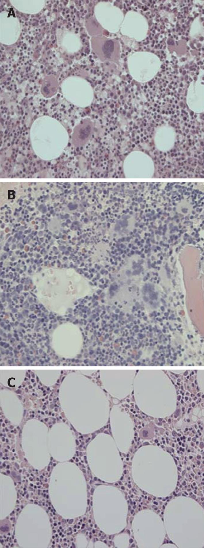
Figure 11 Bone marrow histomorphology from a patient with early prefibrotic polycythemia vera at diagnosis in 1996 (A) (hemoglobin 19.
7 g/dL, hematoctit 0.60. leukocytes normal, platelets 750 × 109/L, modest splenomegaly, JAKV617F > 54%) just prior to interferon α-2b treatment (B) showing typical hypercellular polycythemia vera pictures with clustered large pleiomorphic megakaryocytes and normal cellular bone marrow with megakaryocytes of normal size and morphology (JAK2V617F < 1%) after 8 years of treatment with interferon α-2b treatment (C). Courtesy of Hans Hasselbalch, Denmark. A and B: Pleiomorphic (normal sized and enlarged ) loosely clustered megakaryocytes with hyperploid nuclei and increased cellularity due to increased erythropoiesis in a case of polycythemia vera (PV) before start of interferon α-2b treatment in 1996; C: Megakaryocytes of normal size and morphology in a normocellular bone marrow in the case of PV (A, B) in complete hematological and molecular remission. Originated from Larsen et al[98].
- Citation: Michiels JJ, Ten Kate FW, Koudstaal PJ, Van Genderen PJ. Aspirin responsive platelet thrombophilia in essential thrombocythemia and polycythemia vera. World J Hematol 2013; 2(2): 20-43
- URL: https://www.wjgnet.com/2218-6204/full/v2/i2/20.htm
- DOI: https://dx.doi.org/10.5315/wjh.v2.i2.20


