Copyright
©The Author(s) 2021.
World J Clin Cases. Feb 16, 2021; 9(5): 1221-1227
Published online Feb 16, 2021. doi: 10.12998/wjcc.v9.i5.1221
Published online Feb 16, 2021. doi: 10.12998/wjcc.v9.i5.1221
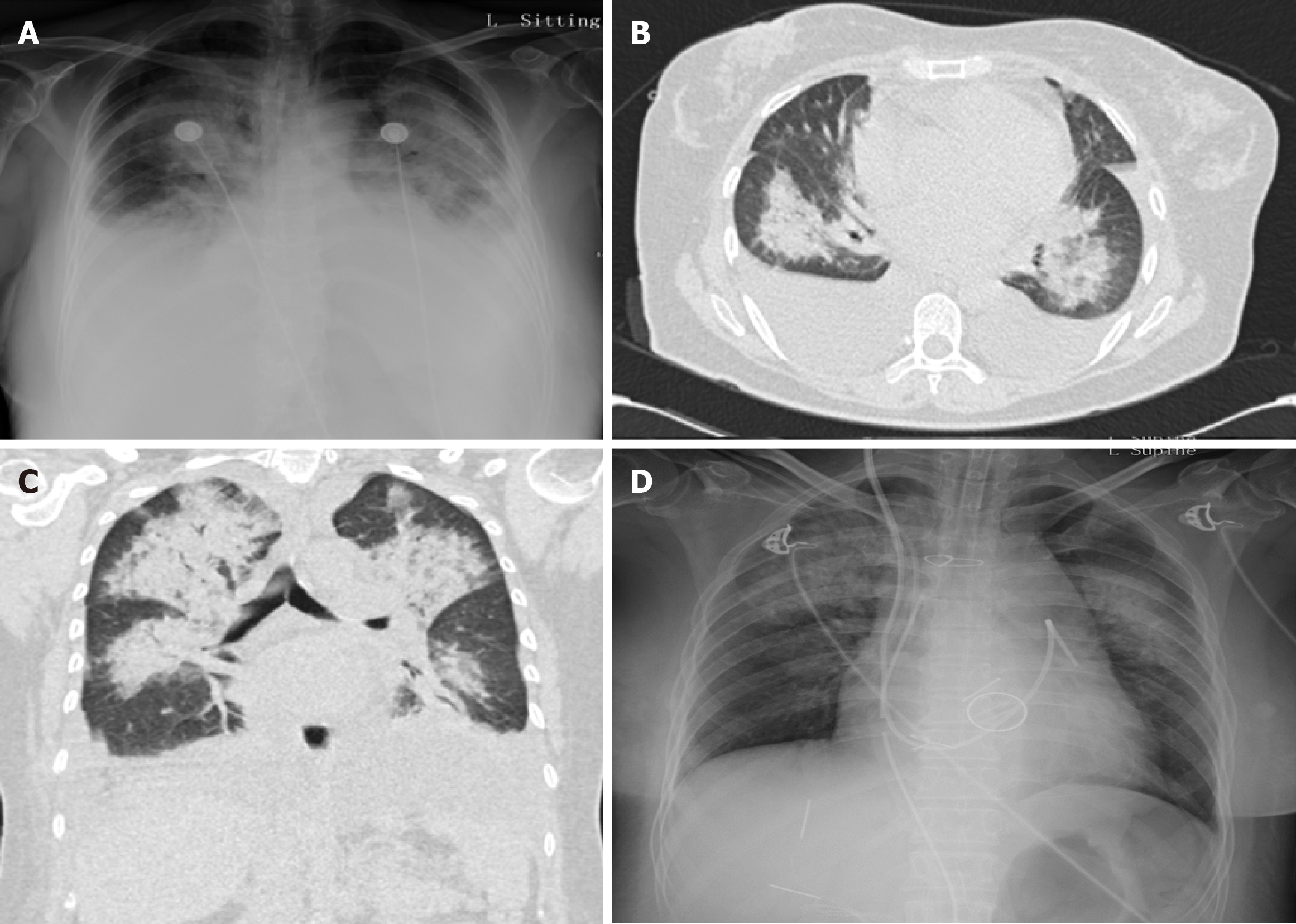
Figure 1 Chest X-ray and computed tomography images.
A: Chest X-ray upon presentation showed diffuse pulmonary edema and bilateral pleural effusion; B and C: Extensive pulmonary edema and bilateral pleural effusion were also observed on computed tomography scanning; D: Chest X-ray showed significant amelioration of pulmonary edema and pleural effusion following replacement of the infected valve.
- Citation: Hou C, Wang WC, Chen H, Zhang YY, Wang WM. Infective bicuspid aortic valve endocarditis causing acute severe regurgitation and heart failure: A case report. World J Clin Cases 2021; 9(5): 1221-1227
- URL: https://www.wjgnet.com/2307-8960/full/v9/i5/1221.htm
- DOI: https://dx.doi.org/10.12998/wjcc.v9.i5.1221


