Copyright
©The Author(s) 2021.
World J Clin Cases. Dec 26, 2021; 9(36): 11406-11418
Published online Dec 26, 2021. doi: 10.12998/wjcc.v9.i36.11406
Published online Dec 26, 2021. doi: 10.12998/wjcc.v9.i36.11406
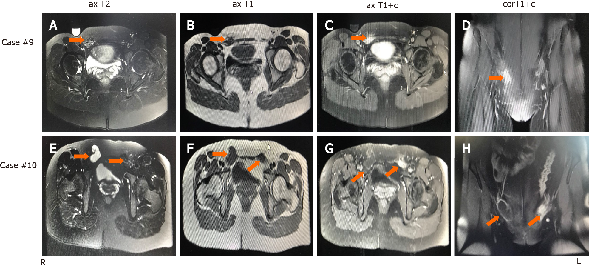
Figure 1 Magnetic resonance imaging findings of inguinal endometriosis.
Magnetic resonance imaging findings of Case 9 (A–D) and Case 10 (E–H). A: Fat-suppressed T2-weighted axial image shows a mixed hyperintense and hypointense lesion with irregular edges in the shape of a cluster of dots and foci in the right groin; B: T1-weighed axial image shows a mixed hyperintense and hypointense lesion in the right groin; C: T1-weighted contrast-enhanced axial image shows an enhanced hyperintense lesion in the right groin; D: T1-weighted contrast-enhanced coronal image shows an enhanced hyperintense lesion in the right groin; E: Fat-suppressed T2-weighted axial image shows a hyperintense cyst in the right groin and a mixed hyperintense and hypointense lesion with irregular edges in the shape of a cluster of dots and foci in the left groin; F: T1-weighed axial image shows hyperintense nodules in part of the wall of the right hernia sac and a mixed hypointense and hyperintense lesion in the left groin; G: T1-weighted contrast-enhanced axial image shows an enhanced hyperintense lesion in part of the thickened wall of the right hernia sac and an enhanced hyperintense lesion in the left groin; H: T1-weighted contrast-enhanced coronal image shows an enhanced hyperintense nodule in the wall of the right hernia sac and an enhanced hyperintense lesion in the left groin.
- Citation: Li SH, Sun HZ, Li WH, Wang SZ. Inguinal endometriosis: Ten case reports and review of literature. World J Clin Cases 2021; 9(36): 11406-11418
- URL: https://www.wjgnet.com/2307-8960/full/v9/i36/11406.htm
- DOI: https://dx.doi.org/10.12998/wjcc.v9.i36.11406


