Published online Dec 6, 2021. doi: 10.12998/wjcc.v9.i34.10666
Peer-review started: April 6, 2021
First decision: July 5, 2021
Revised: July 12, 2021
Accepted: October 25, 2021
Article in press: October 25, 2021
Published online: December 6, 2021
Processing time: 238 Days and 2.1 Hours
Percutaneous coronary intervention can be challenging for ostial coronary artery lesions due to calcium burden and elastic fiber content. Excimer laser coronary atherectomy (ELCA) is a less common treatment for severe calcified coronary ostium lesions.
An 81-year-old male presented to the Cardiology Department of Qingdao Municipal Hospital with a 1-year history of chest pain. Coronary angiography showed severe calcific stenosis (approximately 90%) in the right coronary artery ostium. The right coronary artery ostium was unable to be advanced using a 2.5 mm × 12.0 mm balloon (NC Sprinter, Medtronic, United States) or dilated using a 2.0 mm × 12.0 mm balloon (Sprinter, Medtronic, United States). The patient underwent successful ELCA and balloon dilation of the calcified coronary ostium lesion.
ELCA appears to be a safe and effective treatment for the management of severe calcified coronary ostium lesions.
Core Tip: In the presented case, coronary angiography showed severe calcific stenosis (approximately 90%) in the right coronary artery ostium. A 2.5 mm × 12.0 mm balloon was unable to be advanced into the lesion, while a 2.0 mm × 12.0 mm balloon could not be inflated in the right ostium. Intravascular ultrasonography revealed severe calcifications. The patient underwent an excimer laser coronary atherectomy (ELCA) and balloon dilation, and remained asymptomatic during the 12-mo follow-up. This is the first case report of the successful use of ELCA and small balloon dilatation in treating a severely calcified cardiac ostium lesion.
- Citation: Hou FJ, Ma XT, Zhou YJ, Guan J. Excimer laser coronary atherectomy for a severe calcified coronary ostium lesion: A case report. World J Clin Cases 2021; 9(34): 10666-10670
- URL: https://www.wjgnet.com/2307-8960/full/v9/i34/10666.htm
- DOI: https://dx.doi.org/10.12998/wjcc.v9.i34.10666
Excimer laser coronary atherectomy (ELCA) has emerged as a key procedure that can modify coronary plaques. ELCA achieves its therapeutic efficacy primarily through its photochemical, photothermal, and photomechanical actions. It was reported by Phillips that there were approximately 50000 ELCA catheters used during the period of 2010–2019[1]. However, reports on the use of ELCA in treating heavily calcified coronary lesions are scarce.
An 81-year-old male presented to the Cardiology Department of Qingdao Municipal Hospital with a 1-year history of chest pain.
Coronary angiography (CAG) carried out in a separate center three months earlier noted severe calcific stenosis(approximately 90%) in the right coronary artery (RCA) ostium, 90% stenosis in the proximal left circumflex artery, and 90% stenosis in the proximal left anterior descending artery. The patient declined coronary artery bypass grafting. Stent insertion was then performed in each of the occluded arteries. However, the RCA ostium was unable to be advanced using a 2.5 mm × 12.0 mm balloon (NC Sprinter, Medtronic, United States) or dilated using a 2.0 mm × 12.0 mm balloon (Sprinter, Medtronic, United States). The patient still had persistent angina pectoris despite the insertion of two stents.
The patient had hypertension for more than 30 years, diabetes mellitus for over 7 years, and had no history of smoking.
The patient had no personal or family history.
No positive signs were found during the physical examination.
No abnormalities were found during laboratory examinations.
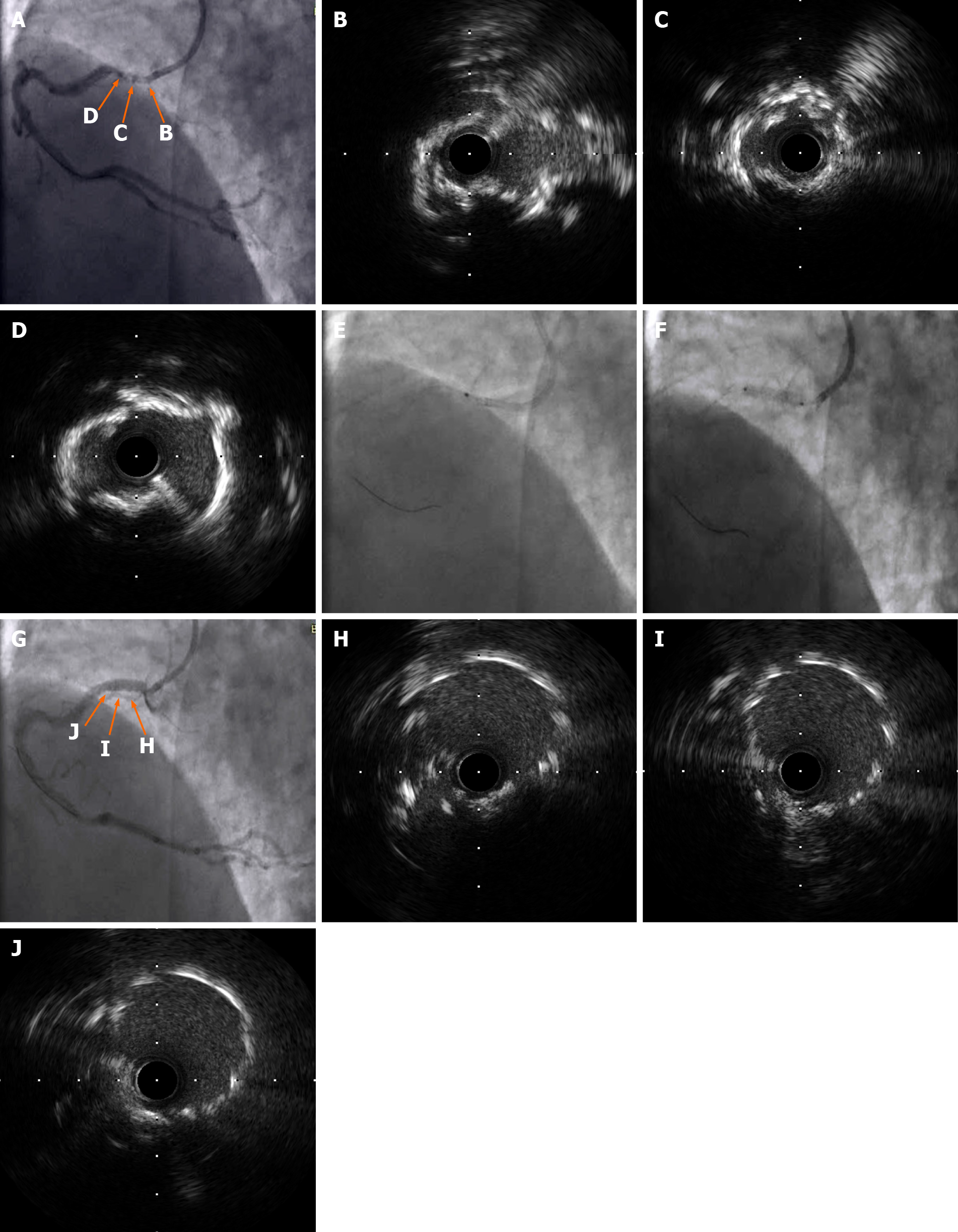
Percutaneous coronary intervention was performed for the RCA ostium lesion after informed consent was obtained from the patient. The radial artery was cannulated using a 6-Fr SAL1.0 guiding catheter (Medtronic, United States). The distal RCA was then cannulated with a Balance Middle Weight Universal II guidewire (Abbott, United States). Intravascular ultrasonography (IVUS; Boston Scientific, United States) was performed and identified severe calcifications (Figure 1A-D).
CAG and IVUS revealed severe calcific stenosis in the RCA ostium.
A 0.9 mm eccentric catheter (Spectranetics, United States) was used to initiate ELCA at 45/60, 60/80, and 80/80 (fluence/Hz) in sequence. This initially resulted in no progress. A 1.5 mm × 15.0 mm balloon (Sprinter, Medtronic, United States) was adopted to dilate the lesion at 10–12 atm. At 45/60 (fluence/Hz), the catheter was slowly passed through the lesion (Figure 1E). The laser catheter was inserted at a speed of less than 0.5-1.0 mm/s using a “saline flush” technique. Normal saline was flushed into the guiding catheter and manifold before laser treatment, and a 5-10 mL bolus of saline was injected prior to each train of laser pulses through the guiding catheter. Continuous saline flushing at a rate of 1-2 mL/s was performed during laser treatment[2,3]. The lesion was then successfully dilated using a 2.5 mm × 12 mm balloon (NC Sprinter, Medtronic, United States). One 3.50 mm × 18.00 mm stent (Xience Xpedition; Abbott) was placed at 12 atm (Figure 1F). The stent was deployed at 14-20 atm to perform post-dilation with a 3.5 mm × 12.0 mm balloon. Stent placements were evaluated with CAG, and the final IVUS findings demonstrated no obvious dissection, malposition, or under expansion (Figure 1G-J).
The patient was healthy and asymptomatic after operation during hospitalization and remained asymptomatic during the 1, 3, 6, and 12-mo follow-up visits.
Coronary artery ostial lesions are barriers to the percutaneous coronary interventions, especially in the presence of severe calcifications. In most cases, severe coronary artery calcifications cannot be crossed or expanded with balloons despite the successful advancement of the guidewire distal to the lesion. ELCA and rotational atherectomy are the two treatments that are effective in managing severe coronary artery calcifications. The risk of no reflow is very low because most of the particles produced by ELCA are less than 10 μm in diameter, which can easily be filtered by the reticuloendothelial system[4]. The dissection of ostium lesions is likely to cause more serious consequences. The use of 308 nm pulsed ultraviolet light reduces the risk of vessel perforation and dissection, given its shallow penetration depth. Most standard 0.014-inch guidewires are compatible with ELCA. Lesions that are unable to be cannulated or expanded may benefit from the use of a 0.9 mm X-80 catheter with a maximum fluence (energy) of 80 mJ/mm2 and repetition rate of 80 Hz, both of which are attainable using a 10 s on and 5 s off laser cycle[1]. Calcified lesions are amenable to treatment with a 0.9 mm excimer laser catheter bringing increased density of energy while conserving the production of heat, which results in a smaller ablated area. The laser catheter pushing speed should be less than 0.5-1.0 mm/s to avoid production of large particles.
Currently, ELCA is commonly used in highly complex lesions, including saphenous vein grafts, calcifications, tortuosity (moderate/severe), in-stent restenosis, and bifurcations, and carries the dual benefit of low rates of major adverse cardiovascular complications and high technical and procedural success rates[5,6]. We present the first case of a severe calcified ostium lesion treated with ELCA and balloon dilatation. During the ELCA procedure, if the catheter cannot pass through the lesion smoothly, balloon dilatation can be used to change the plaque morphology and achieve better results. Although ELCA may not be the first choice for severe calcified coronary ostium lesions as compared to rotational atherectomy, it may be used as an alternative in the following cases: Thrombus, severe tortuosity, bifurcation, ostial coronary artery dissection, the failure of a Rotawire to pass through the target lesion, and severe heart failure. We successfully treated a severe calcified coronary ostium lesion by ELCA and small balloon dilatation. Nevertheless, further studies are needed to evaluate the efficacy and safety of ELCA vs rotational atherectomy.
Alternative use of ELCA and small balloon dilatation appears to be a safe and effective means of managing severely calcified coronary ostium lesions.
Provenance and peer review: Unsolicited article; Externally peer reviewed.
Specialty type: Medicine, research and experimental
Country/Territory of origin: China
Peer-review report’s scientific quality classification
Grade A (Excellent): 0
Grade B (Very good): 0
Grade C (Good): C
Grade D (Fair): 0
Grade E (Poor): 0
P-Reviewer: Matsuo Y S-Editor: Fan JR L-Editor: A P-Editor: Fan JR
| 1. | Tsutsui RS, Sammour Y, Kalra A, Reed G, Krishnaswamy A, Ellis S, Nair R, Khatri J, Kapadia S, Puri R. Excimer Laser Atherectomy in Percutaneous Coronary Intervention: A Contemporary Review. Cardiovasc Revasc Med. 2021;25:75-85. [RCA] [PubMed] [DOI] [Full Text] [Cited by in Crossref: 9] [Cited by in RCA: 39] [Article Influence: 7.8] [Reference Citation Analysis (0)] |
| 2. | Shavadia JS, Vo MN, Bainey KR. Challenges With Severe Coronary Artery Calcification in Percutaneous Coronary Intervention: A Narrative Review of Therapeutic Options. Can J Cardiol. 2018;34:1564-1572. [RCA] [PubMed] [DOI] [Full Text] [Cited by in Crossref: 17] [Cited by in RCA: 24] [Article Influence: 3.4] [Reference Citation Analysis (0)] |
| 3. | Shibui T, Tsuchiyama T, Masuda S, Nagamine S. Excimer laser coronary atherectomy prior to paclitaxel-coated balloon angioplasty for de novo coronary artery lesions. Lasers Med Sci. 2021;36:111-117. [RCA] [PubMed] [DOI] [Full Text] [Full Text (PDF)] [Cited by in Crossref: 4] [Cited by in RCA: 10] [Article Influence: 2.0] [Reference Citation Analysis (0)] |
| 4. | Rawlins J, Din JN, Talwar S, O'Kane P. Coronary Intervention with the Excimer Laser: Review of the Technology and Outcome Data. Interv Cardiol. 2016;11:27-32. [RCA] [PubMed] [DOI] [Full Text] [Cited by in Crossref: 36] [Cited by in RCA: 58] [Article Influence: 6.4] [Reference Citation Analysis (0)] |
| 5. | Karacsonyi J, Armstrong EJ, Truong HTD, Tsuda R, Kokkinidis DG, Martinez-Parachini JR, Alame AJ, Danek BA, Karatasakis A, Roesle M, Khalili H, Ungi I, Banerjee S, Brilakis ES, Rangan BV. Contemporary Use of Laser During Percutaneous Coronary Interventions: Insights from the Laser Veterans Affairs (LAVA) Multicenter Registry. J Invasive Cardiol. 2018;30:195-201. [PubMed] |
| 6. | Ojeda S, Azzalini L, Suárez de Lezo J, Johal GS, González R, Barman N, Hidalgo F, Bellera N, Dangas G, Jurado-Román A, Kini A, Romero M, Moreno R, Garcia Del Blanco B, Mehran R, Sharma SK, Pan M. Excimer laser coronary atherectomy for uncrossable coronary lesions. A multicenter registry. Catheter Cardiovasc Interv. 2020;. [RCA] [PubMed] [DOI] [Full Text] [Cited by in Crossref: 6] [Cited by in RCA: 19] [Article Influence: 3.8] [Reference Citation Analysis (0)] |


