Published online Nov 6, 2021. doi: 10.12998/wjcc.v9.i31.9564
Peer-review started: March 13, 2021
First decision: August 18, 2021
Revised: August 20, 2021
Accepted: September 14, 2021
Article in press: September 14, 2021
Published online: November 6, 2021
Processing time: 230 Days and 7.8 Hours
Primary soft tissue giant cell tumor (GCT-ST) is rare and has relatively low malignant potential. Most reports are pathological and clinical studies, while imaging studies have only been reported in cases of adjacent bone or with atypical cystic degeneration. With regard to the findings on magnetic resonance imaging (MRI) or ultrasonography, superficial masses can be further identified based on facial edema, skin thickening, skin contact, internal hemorrhage or necrosis and lobulation of the mass. Unlike deep-seated masses, MRI features do not always provide an accurate diagnosis for benign and malignant patients with superficial soft-tissue lesions. Thus, the application of diffusion-weighted imaging (DWI) to evaluate superficial soft tissue tumors is necessary.
A 36-year-old woman who had a suspected malignant tumor in the upper limb on ultrasound and computed tomography is reported. The signal intensity of the suspected tumor was heterogeneous on plain MRI; nodular and heterogeneous enhancement was observed in the tumor with irregular shapes and blurred margins on dynamic contrast-enhanced MRI. The lesion on DWI was hyperintense with a higher mean apparent diffusion coefficient (ADC) value. Finally, a GCT-ST was confirmed by pathology. This case suggests that GCT-ST should be distinguished as a benign soft tissue mass from giant cell-rich soft tissue neoplasms or malignant tumors.
The MRI features of the superficial GCT-ST in the upper limb included heterogeneous signal intensity within the lesion on T2-weighted image (T2WI) and T1-weighted fat-saturation spoiled gradient recalled echo (T1 FSPGR), nodular enhancement with blurred margins, irregular shapes, and a slow-increased enhancement. DWI could be used to differentiate a benign soft tissue mass from a malignant mass by the mean ADC value and provide more radiologic-pathologic information for the diagnosis of GCT-ST. Comprehensive imaging of primary GCT-ST could help complete tumor resection, and in turn likely prolong survival after surgery.
Core Tip: The comprehensive magnetic resonance imaging (MRI) features of primary superficial soft tissue giant cell tumor (GCT-ST) in the upper limb were reported in our case. The manifestations of MRI included heterogeneous signal intensity within the lesion on T2-weighted image and T1-weighted fat-saturation spoiled gradient recalled echo, nodular enhancement with blurred margins, irregular shapes, and a slow-increased enhancement. Diffusion-weighted imaging could be used in the differential diagnosis by the mean apparent diffusion coefficient value and provide more radiologic-pathologic information for the diagnosis of GCT-ST.
- Citation: Kang JY, Zhang K, Liu AL, Wang HL, Zhang LN, Liu WV. Characteristics of primary giant cell tumor in soft tissue on magnetic resonance imaging: A case report. World J Clin Cases 2021; 9(31): 9564-9570
- URL: https://www.wjgnet.com/2307-8960/full/v9/i31/9564.htm
- DOI: https://dx.doi.org/10.12998/wjcc.v9.i31.9564
Primary giant cell tumor of soft tissue (GCT-ST) is rare and usually located in superficial and deep soft tissues, and has relatively low malignant potential[1]. Histologically, GCT-ST lesions bear a close resemblance to their bony counterparts, giant cell tumor of bone. Most existing reports are pathological and clinical studies, while imaging studies have only been reported in cases of adjacent bone or with atypical cystic degeneration[2-11]. With regard to the findings on magnetic resonance imaging (MRI) or ultrasonography, superficial masses in the soft tissue can be further identified based on facial edema, skin thickening, skin contact, internal hemorrhage or necrosis and lobulation of the mass[12-15].
Unlike patients with deep-seated masses, size (i.e., 50 mm in diameter) is not an important factor in superficial soft-tissue lesions. However, MRI features do not always provide an accurate diagnosis for benign and malignant soft tissue lesions. The application of diffusion-weighted imaging (DWI) to differentiate benign from malignant soft tissue tumors is necessary. So far, only one case was reported on the juxtacortical mass of GCT-ST lesions using intravoxel incoherent motion (IVIM) DWI[2]. Moreover, qualitative and quantitative data from conventional MRI can improve the diagnosis of benign and malignant soft-tissue masses[11]. Therefore, the aims of the current study were to report the case of GCT-ST in the upper limb using comprehensive medical imaging examinations especially quantitative DWI and to present a literature review on the topic.
A 36-year-old woman was admitted to our hospital with swelling, skin redness and pain in the upper limb that persisted for 6 mo without a prior history of trauma.
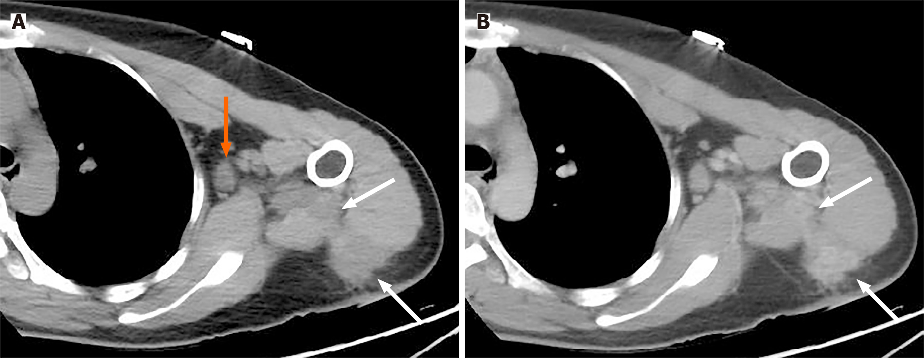
The patient underwent ultrasound (US) and a computed tomography (CT) scan. The US revealed a blurred large solid mass in the deltoid muscle. Plain CT showed a hypodense mass in the superficial deltoid muscle extending to the intermuscular space (Figure 1A, white arrow) with axillary lymphadenopathy (Figure 1A, orange arrow), while contrast-enhanced CT revealed a slightly heterogeneous enhancement of the mass with blurred margins (Figure 1B, white arrow). There was no evidence of calcification or mineralization in the mass, and the adjacent structures were all normal.
A clinical examination revealed that the 6.0 cm x 4.0 cm mass was tender without discharge or drainage sinus.
MRI: MRI was performed using a 1.5T whole-body MR scanner (Signa, Excite, HDxt, General Electric Healthcare, Milwaukee, WI, USA). The MRI protocols were as follows (Table 1): (1) Conventional MR scan sequences included coronal or axial T2-weighted fast spin-echo (FSE) images, T1-weighted fat-saturation spoiled gradient recalled echo (T1 FSPGR) images; (2) Axial T1 3D FSPGR liver acquisition with volume acceleration (LAVA) sequence (total 4 phases, repetition time/echo time (TR/TE) = 6.0 ms/3.0 ms, FA = 12º, slice thickness/slice spacing = 5.0 mm/2.5 mm, acquisition time = 23 s/one phase) was performed after an injection of 0.1 mmol/kg gadolinium using an antecubital vein power injector at a rate of 2.0 mL/s followed by 20 mL saline; the first acquisition started 25 s after contrast agent injection; and (3) DWI (b = 500 s/mm2) with TR/TE of 4700 ms/69 ms, slice thickness/slice spacing of 6.0 mm/1.5 mm, a field of view (FOV) of 280 mm x 252 mm and matrix size of 256 × 256 were used; DWI was performed before contrast injection.
| Scan sequence | TR (ms) | TE (ms) | FOV (mm) | Thickness (mm) | Gap (mm) | Flip angles | NEX | Matrix (mm) |
| T1 FSPGR | 235 | 3.2 | 280 x 224 | 6.0 | 1.5 | 80º | 3 | 256 × 192 |
| T2WI | 3940 | 87 | 260 x 208 | 5.0 | 1.0 | 90º | 3 | 320 × 192 |
| DWI | 4700 | 69 | 280 x 252 | 6.0 | 1.5 | 90º | 4 | 128 × 128 |
| LAVA | 5.8 | 3.1 | 280 x 224 | 5.0 | -2.5 | 12° | 1 | 224 × 160 |
MRI analysis: All MR images were transferred to a GE workstation (Advantage Windows 4.5; General Electric, Madison, WI, USA) for image processing and were interpreted by two radiologists with more than five years of diagnostic experience. Only the consensus of any diagnosis between the two readers was used for the final MRI analysis. The following lesion characteristics were recorded: (1) Signal intensity on T2WI and T1 FSPGR; (2) Morphology and maximum lesion size on dynamic contrast-enhanced MRI (DCE-MRI); (3) Time-intensity curve (TIC) obtained from DCE-MRI; and (4) Apparent diffusion coefficient (ADC) value obtained from DWI.
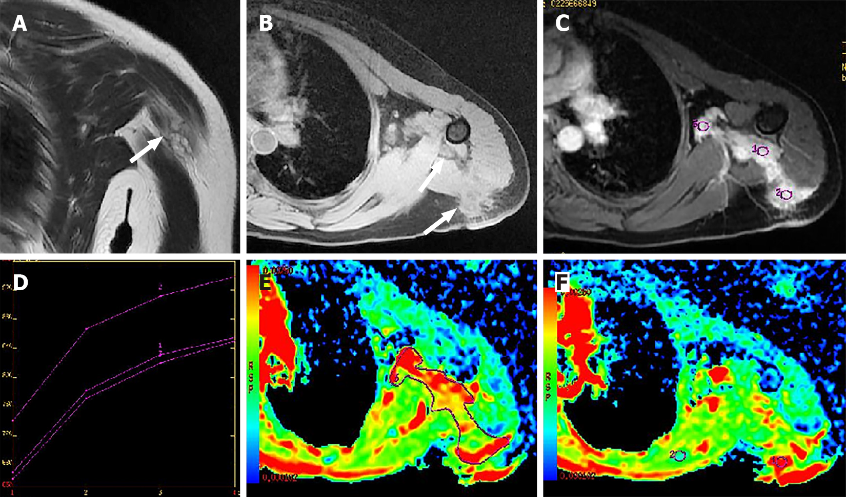
On morphological MR images, the manifestations of the mass were obvious. The maximum lesion size was 6.0 cm. Compared to muscle signal intensity, the lesion showed hyper-intensity on T2WI without fat saturation (Figure 2A, white arrow), and heterogeneous hypo-intensity to iso-intensity on T1 FSPGR (Figure 2B, white arrow). On DCE-MRI, the solid mass showed multiple nodular enhancements with irregular shapes and blurred margins (Figure 2C). Furthermore, internal enhancement was heterogeneous. The TIC showed slow increased type (Figure 2D). On DWI (b = 500 s/mm2), the lesion was hyperintense with a higher mean ADC value of 2.19 × 10⁻³ mm2/s (Figure 2E, ROI 3) than surrounding normal soft tissue (1.03 × 10⁻³ mm2/s) (Figure 2F, ROI 2).
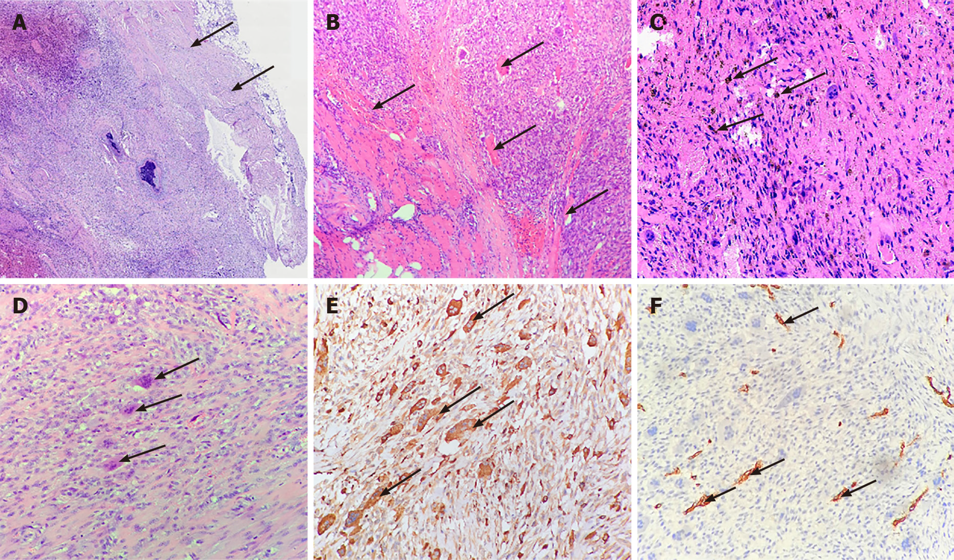
The resected specimen revealed that the tumor invaded the peripheral muscles. Consequently, complete surgical excision of the mass was performed. Pathological examination indicated that many multinucleated giant cells were scattered and surrounded by spindle cells, suggesting a GCT-ST. The giant and spindle cells had slight cellular atypia. In addition, spindle cells exhibited brisk mitotic activity. The tumors invaded the neighboring muscles. The obtained samples showed a slow-growing entity.
Tumor interstitial hemorrhage was obvious and rich in hemosiderin-containing cells (Figure 3A-D). Immunohistochemically, the giant cells were strongly positive for CD68, no staining of myoglobin, myogenin, MyoD1, desmin, CD34, S-100, CK, and EMA were found in the tumor (Figure 3E, F). The patient was finally diagnosed with giant cell tumors based on clinical, histologic and immunohistochemical (IHC) findings.
Complete surgical excision of the mass was performed.
No metastasis or recurrence was found in this case on 1-year MRI follow-up.
The diagnosis of GCT-ST based on preoperative US and CT can be challenging due to non-specific manifestations and difficulty in defining tumor extent. In our study, we used conventional MRI and DWI to investigate a case of GCT-ST in the upper limb. The lesion abnormalities were limited to soft tissues without obvious calcification or bone erosion as frequently observed at the periphery of GCT-ST tumors[2-7]. In addition, the lesion on MRI presented with a solid mass and blurry upper and lower boundaries. There was slight hyper-intensity on T2WI, indicating that the tumor invaded the neighboring muscles. Thus, our results suggested that MRI-DWI can be useful in identifying soft-tissue masses with no malignant potential. In our study, the lesion showed a non-homogeneous hypo-intensity to iso-intensity on T1WI and hyper-intensity on T2WI. Consistent with previous reports, MR images of bone GCT may suggest the presence of solid components with hypo-intensity to iso-intensity on T1WI and T2WI, caused by hemosiderin deposition or high collagen content[3,4]. On contrast-enhanced images, unlike malignant soft tissue tumors or GCT-ST tumors with diffuse cystic components located in the subcutaneous tissue[8-11], the solid region of the lesion was slowly enhanced like hypervascular tissue, in line with IHC performance such as in bone GCT and the findings in other benign soft tissue tumors[2-4].
In the present study, the lesion was significantly large (maximum lesion size was 6.0 cm) and located in the superficial areas of the extremity or trunk. This type of lesion is commonly observed in GCT-ST, but it can also be found in the deep soft tissues of the head, neck, and retroperitoneum region[2-10]. Unlike deep-seated masses, size (i.e., 5 cm in diameter) is not an essential indicative factor of malignancy for superficial masses[12]. To determine the benign characteristics of GCT-ST on MRI, we attempted to evaluate the feasibility of DWI.
DWI is used to differentiate benign from malignant soft tissue lesions based on ADC values as quantitative DWI is useful for differentiating between malignant and benign superficial masses[12-14]. Typically, if the ADC value is higher than the threshold of 1.0 × 10⁻³ mm2/s, the superficial soft-tissue mass is considered to be benign[12]. In this study, DW images were acquired with a b value of 500 s/mm2 and the mean ADC value of solid GCT-ST lesions was 2.19 × 10⁻³ mm2/s, which was higher than the surrounding healthy tissue and its threshold. The reason for an overlap between the ADC values in benign and malignant tumors might be the heterogeneous nature of soft-tissue tumors such as intratumoral water content that increases diffusion, mucinous contents and intratumoral necrosis[12,13]. We speculated that high ADC values of the solid GCT-ST were consistent with tumor histology as intensely enhanced solid masses with no detectable macroscopic necrotic or myxoid predominant areas despite tumor interstitial hemorrhage were observed. Future studies with a higher sample size are needed to further confirm that a high ADC value is a characteristic of GCT-ST.
Clinically, primary GCT-ST mainly affects young to middle-aged adults[4-11,16]. Here, our patient was a young woman who had an ill-circumscribed multinodular mass covered by a fleshy red-brown surface and pain in the upper limb. Although the lesion in the upper limb had axillary lymphadenopathy and infiltrated adjacent anatomical structures, including neurovascular bundles, the lesion was unilateral and identified as a benign GCT-ST.
A noncalcified or unossified soft-tissue mass with low signal on T2WI is usually fibrous with little cellularity. As the observed mass with solid components in our study was not a bone lesion, giant cell tumors of tendon sheath (GCT-TS) and giant cell-rich forms of nodular fasciitis (NF) were included in the differential diagnosis. A GCT-TS is generally located near joint spaces, while a cystic lesion and metaplastic bone formation are absent, and calcification or ossification are very rare. GCT-TS always shows hyper-intensity on contrast-enhanced images[17,18]. NF is usually located in the subcutaneous tissue and shows hyper-intensity on T2WI caused by fluid-filled mucoid spaces. NF on MR images is more variable. Differential diagnosis of these giant cell-rich soft tissue neoplasms is important as clinical behavior, prognosis, and treatment can significantly differ[15-22].
Our case suggested that the MRI features of superficial GCT-STs in the upper limb, including heterogeneous signal intensity within the lesion on T2WI and T1 FSPGR, nodular enhancement with blurred margins, irregular shapes, and a slow enhancement of TIC on DCE-MRI. In addition, DWI could be used to differentiate a benign soft tissue mass from the malignant one by the mean ADC value, thus providing more radiologic-pathologic information for the diagnosis of GCT-ST. Comprehensive imaging of primary GCT-ST can aid in complete tumor resection, which in turn might promote long-term survival after surgery. However, these findings need to be confirmed using more samples.
Manuscript source: Unsolicited manuscript
Specialty type: Radiology, nuclear medicine and medical imaging
Country/Territory of origin: China
Peer-review report’s scientific quality classification
Grade A (Excellent): 0
Grade B (Very good): 0
Grade C (Good): C
Grade D (Fair): 0
Grade E (Poor): 0
P-Reviewer: Muthu S S-Editor: Ma YJ L-Editor: Webster JR P-Editor: Guo X
| 1. | Jo VY, Fletcher CD. WHO classification of soft tissue tumours: an update based on the 2013 (4th) edition. Pathology. 2014;46:95-104. [RCA] [PubMed] [DOI] [Full Text] [Cited by in Crossref: 505] [Cited by in RCA: 671] [Article Influence: 61.0] [Reference Citation Analysis (0)] |
| 2. | Lee MY, Jee WH, Jung CK, Yoo IeR, Chung YG. Giant cell tumor of soft tissue: a case report with emphasis on MR imaging. Skeletal Radiol. 2015;44:1039-1043. [RCA] [PubMed] [DOI] [Full Text] [Cited by in Crossref: 10] [Cited by in RCA: 9] [Article Influence: 0.9] [Reference Citation Analysis (0)] |
| 3. | Chen L, Shi XL, Zhou ZM, Qin LD, Liu XH, Jiang L, Zhang QJ, Ding XY. Clinical Significance of MRI and Pathological Features of Giant Cell Tumor of Bone Boundary. Orthop Surg. 2019;11:628-634. [RCA] [PubMed] [DOI] [Full Text] [Full Text (PDF)] [Cited by in Crossref: 4] [Cited by in RCA: 5] [Article Influence: 0.8] [Reference Citation Analysis (1)] |
| 4. | Murphey MD, Nomikos GC, Flemming DJ, Gannon FH, Temple HT, Kransdorf MJ. From the archives of AFIP. Imaging of giant cell tumor and giant cell reparative granuloma of bone: radiologic-pathologic correlation. Radiographics. 2001;21:1283-1309. [RCA] [PubMed] [DOI] [Full Text] [Cited by in Crossref: 353] [Cited by in RCA: 294] [Article Influence: 12.3] [Reference Citation Analysis (0)] |
| 5. | Allen PW. Primary giant cell tumors of soft tissue. Am J Surg Pathol. 2000;24:1175-1176. [RCA] [PubMed] [DOI] [Full Text] [Cited by in Crossref: 2] [Cited by in RCA: 2] [Article Influence: 0.1] [Reference Citation Analysis (0)] |
| 6. | Sonmez Ergun S, Buyukbabani N, Atilganoglu U. Primary giant cell tumor of soft tissue mimicking a vascular neoplasm. Dermatol Surg. 2008;34:102-104. [RCA] [PubMed] [DOI] [Full Text] [Cited by in Crossref: 2] [Cited by in RCA: 2] [Article Influence: 0.1] [Reference Citation Analysis (0)] |
| 7. | Callı AO, Tunakan M, Katilmiş H, Kilçiksiz S, Oztürkcan S. Soft tissue giant cell tumor of low malignant potential of the neck: a case report and review of the literature. Turk Patoloji Derg. 2014;30:73-77. [RCA] [PubMed] [DOI] [Full Text] [Cited by in Crossref: 1] [Cited by in RCA: 4] [Article Influence: 0.4] [Reference Citation Analysis (0)] |
| 8. | Kishi S, Monma H, Hori H, Kinugasa S, Fujimoto M, Nakamura T. First Case Report of a Huge Giant Cell Tumor of Soft Tissue Originating from the Retroperitoneum. Am J Case Rep. 2018;19:642-650. [RCA] [PubMed] [DOI] [Full Text] [Full Text (PDF)] [Cited by in Crossref: 3] [Cited by in RCA: 3] [Article Influence: 0.4] [Reference Citation Analysis (0)] |
| 9. | An SB, Choi JA, Chung JH, Oh JH, Kang HS. Giant cell tumor of soft tissue: a case with atypical US and MRI findings. Korean J Radiol. 2008;9:462-465. [RCA] [PubMed] [DOI] [Full Text] [Full Text (PDF)] [Cited by in Crossref: 17] [Cited by in RCA: 20] [Article Influence: 1.3] [Reference Citation Analysis (0)] |
| 10. | Boneschi V, Parafioriti A, Armiraglio E, Gaiani F, Brambilla L. Primary giant cell tumor of soft tissue of the groin - a case of 46 years duration. J Cutan Pathol. 2009;36 Suppl 1:20-24. [RCA] [PubMed] [DOI] [Full Text] [Cited by in Crossref: 8] [Cited by in RCA: 10] [Article Influence: 0.6] [Reference Citation Analysis (0)] |
| 11. | Dodd LG, Major N, Brigman B. Malignant giant cell tumor of soft parts. Skeletal Radiol. 2004;33:295-299. [RCA] [PubMed] [DOI] [Full Text] [Cited by in Crossref: 21] [Cited by in RCA: 21] [Article Influence: 1.0] [Reference Citation Analysis (0)] |
| 12. | Jeon JY, Chung HW, Lee MH, Lee SH, Shin MJ. Usefulness of diffusion-weighted MR imaging for differentiating between benign and malignant superficial soft tissue tumours and tumour-like lesions. Br J Radiol. 2016;89:20150929. [RCA] [PubMed] [DOI] [Full Text] [Cited by in Crossref: 26] [Cited by in RCA: 32] [Article Influence: 3.6] [Reference Citation Analysis (0)] |
| 13. | Choi YJ, Lee IS, Song YS, Kim JI, Choi KU, Song JW. Diagnostic performance of diffusion-weighted (DWI) and dynamic contrast-enhanced (DCE) MRI for the differentiation of benign from malignant soft-tissue tumors. J Magn Reson Imaging. 2019;50:798-809. [RCA] [PubMed] [DOI] [Full Text] [Cited by in Crossref: 24] [Cited by in RCA: 42] [Article Influence: 7.0] [Reference Citation Analysis (0)] |
| 14. | Bonarelli C, Teixeira PA, Hossu G, Meyer JB, Chen B, Gay F, Blum A. Impact of ROI Positioning and Lesion Morphology on Apparent Diffusion Coefficient Analysis for the Differentiation Between Benign and Malignant Nonfatty Soft-Tissue Lesions. AJR Am J Roentgenol. 2015;205:W106-W113. [RCA] [PubMed] [DOI] [Full Text] [Cited by in Crossref: 15] [Cited by in RCA: 18] [Article Influence: 1.8] [Reference Citation Analysis (0)] |
| 15. | Khoo MM, Tyler PA, Saifuddin A, Padhani AR. Diffusion-weighted imaging (DWI) in musculoskeletal MRI: a critical review. Skeletal Radiol. 2011;40:665-681. [RCA] [PubMed] [DOI] [Full Text] [Cited by in Crossref: 173] [Cited by in RCA: 165] [Article Influence: 11.8] [Reference Citation Analysis (0)] |
| 16. | Meana Moris AR, García Gonzalez P, Fuente Martin E, Gonzalez Suarez C, Moro Barrero L. Primary giant cell tumor of soft tissue: fluid-fluid levels at MRI (2010:3b). Eur Radiol. 2010;20:1539-1543. [RCA] [PubMed] [DOI] [Full Text] [Cited by in Crossref: 12] [Cited by in RCA: 11] [Article Influence: 0.7] [Reference Citation Analysis (0)] |
| 17. | Hourani R, Taslakian B, Shabb NS, Nassar L, Hourani MH, Moukarbel R, Sabri A, Rizk T. Fibroblastic and myofibroblastic tumors of the head and neck: comprehensive imaging-based review with pathologic correlation. Eur J Radiol. 2015;84:250-260. [RCA] [PubMed] [DOI] [Full Text] [Cited by in Crossref: 21] [Cited by in RCA: 21] [Article Influence: 2.1] [Reference Citation Analysis (0)] |
| 18. | Kwak HS, Lee SY, Kim JR, Lee KB. MR imaging of calcifying aponeurotic fibroma of the thigh. Pediatr Radiol. 2004;34:438-440. [RCA] [PubMed] [DOI] [Full Text] [Cited by in Crossref: 20] [Cited by in RCA: 15] [Article Influence: 0.7] [Reference Citation Analysis (0)] |
| 19. | Wang C, Song RR, Kuang PD, Wang LH, Zhang MM. Giant cell tumor of the tendon sheath: Magnetic resonance imaging findings in 38 patients. Oncol Lett. 2017;13:4459-4462. [RCA] [PubMed] [DOI] [Full Text] [Cited by in Crossref: 23] [Cited by in RCA: 35] [Article Influence: 4.4] [Reference Citation Analysis (0)] |
| 20. | Ward CM, Lueck NE, Steyers CM. Acute carpal tunnel syndrome caused by diffuse giant cell tumor of tendon sheath: a case report. Iowa Orthop J. 2007;27:99-103. [RCA] [PubMed] [DOI] [Full Text] [Cited by in Crossref: 1] [Cited by in RCA: 1] [Article Influence: 0.1] [Reference Citation Analysis (0)] |
| 21. | Wang XL, De Schepper AM, Vanhoenacker F, De Raeve H, Gielen J, Aparisi F, Rausin L, Somville J. Nodular fasciitis: correlation of MRI findings and histopathology. Skeletal Radiol. 2002;31:155-161. [RCA] [PubMed] [DOI] [Full Text] [Cited by in Crossref: 86] [Cited by in RCA: 88] [Article Influence: 3.8] [Reference Citation Analysis (0)] |
| 22. | Nakayama R, Jagannathan JP, Ramaiya N, Ferrone ML, Raut CP, Ready JE, Hornick JL, Wagner AJ. Clinical characteristics and treatment outcomes in six cases of malignant tenosynovial giant cell tumor: initial experience of molecularly targeted therapy. BMC Cancer. 2018;18:1296. [RCA] [PubMed] [DOI] [Full Text] [Full Text (PDF)] [Cited by in Crossref: 11] [Cited by in RCA: 26] [Article Influence: 3.7] [Reference Citation Analysis (0)] |


