Copyright
©The Author(s) 2021.
World J Clin Cases. Oct 6, 2021; 9(28): 8571-8578
Published online Oct 6, 2021. doi: 10.12998/wjcc.v9.i28.8571
Published online Oct 6, 2021. doi: 10.12998/wjcc.v9.i28.8571
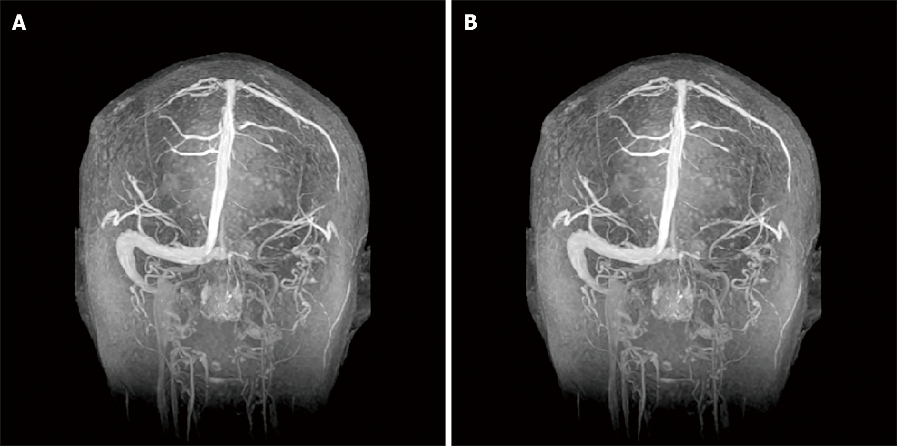
Figure 3 Magnetic resonance venogram.
A: Thrombosis in the left internal jugular vein, transverse sinus, sigmoid sinus, confluent sinus, partial venous thrombosis in the straight sinus and inferior sagittal sinus; B: The thrombus was smaller than before.
- Citation: Song XH, Xu T, Zhao GH. Hypereosinophilia with cerebral venous sinus thrombosis and intracerebral hemorrhage: A case report and review of the literature. World J Clin Cases 2021; 9(28): 8571-8578
- URL: https://www.wjgnet.com/2307-8960/full/v9/i28/8571.htm
- DOI: https://dx.doi.org/10.12998/wjcc.v9.i28.8571


