Published online Sep 6, 2021. doi: 10.12998/wjcc.v9.i25.7535
Peer-review started: February 23, 2021
First decision: April 13, 2021
Revised: April 20, 2021
Accepted: May 20, 2021
Article in press: May 20, 2021
Published online: September 6, 2021
Processing time: 188 Days and 13.5 Hours
The most commonly ingested foreign body in Asians is fish bone. The vast majority of patients have obvious symptoms and can be timely diagnosed and treated. Cases of pyogenic cervical spondylitis and diskitis with retropharyngeal and epidural abscess resulting in incomplete quadriplegia due to foreign body ingestion have been rarely reported. The absence of pharyngeal or esophageal discomfort and negative computed tomography (CT) findings of fish bone have not been reported. We report the case of an elderly female patient with delayed cervical infection and incomplete quadriplegia who had a history of fish bone ingestion.
A 73-year-old woman presented with right neck pain and weakness of four limbs for a week, and had a history of fish bone ingestion and negative findings on laryngoscopic examination one month previously. She did not complain of any pharyngeal or esophageal discomfort. Cervical magnetic resonance imaging showed C4/C5 spondylitis and diskitis along with retropharyngeal and ventral epidural abscesses. No sign of fish bone was detected on lateral cervical radiography and CT scans. The muscle strength of the patient’s right lower limb receded to grade 1 and other limbs to grade 2 suddenly on the 10th day of hospitalization. Emergency surgery was performed to drain the abscess and decompress the spinal cord by removing the anterior inflammatory necrotic tissue. Simultaneously, flexible esophagogastroduodenoscopy was carried out and a hole in the posterior pharyngeal wall was found. The motor weakness of the right lower limb improved to grade 3 and the other limbs to grade 4 within 2 d postoperatively.
This rare case highlights the awareness of the posterior pharyngeal or esophageal wall perforation in patients with cervical pyogenic spondylitis along with a history of fish bone ingestion, even though local discomfort symptoms are absent and the radiological examinations are negative.
Core Tip: We report a rare case of an elderly female patient with delayed cervical infection and incomplete quadriplegia who had a history of fish bone ingestion. We recommend clinicians be aware of this rare condition that can occur without positive computed tomography findings and mediastinitis. In addition, local complete debridement, sufficient use of antibiotics and placement of a gastric tube for nasal feeding played a vital role in infection control and healing of the laryngopharyngeal wall perforation.
- Citation: Li SY, Miao Y, Cheng L, Wang YF, Li ZQ, Liu YB, Zou TM, Shen J. Surgical treatment of delayed cervical infection and incomplete quadriplegia with fish-bone ingestion: A case report. World J Clin Cases 2021; 9(25): 7535-7541
- URL: https://www.wjgnet.com/2307-8960/full/v9/i25/7535.htm
- DOI: https://dx.doi.org/10.12998/wjcc.v9.i25.7535
In Asians, the most commonly ingested foreign body is fish bone[1]. The vast majority of patients have obvious symptoms and can be timely diagnosed and treated[2,3]. Few cases may develop pharyngeal or esophageal wall perforation[4]. As far as we know, this is the first reported case of an ingested fish-bone with no obvious clinical manifestation found until the occurrence of severe cervical infection of C4/C5 and incomplete quadriplegia one month later.
A 73-year-old woman was referred to our hospital due to right neck pain and weakness of all four limbs for one week, urine retention and occasional high fever for three days.
She had a history of fish bone ingestion one month ago, and laryngoscopic examination did not show positive findings. The patient did not complain of any pharyngeal or esophageal discomfort.
She had an 8-year history of diabetes and poor glycemic control.
The patient had unremarkable personal and familial medical history, including psycho-social history.
On admission, her body temperature was 36.5℃. Physical examination showed sensitive percussion over the cervical spinous processes and marked restriction of neck motion. Lhermitte’s sign was positive. The strength of the right upper and lower extremities was grade 3 and of the left upper and lower extremities was grade 4. No sensory deficit was observed. The bilateral pathologic reflexes were positive.
The muscle strength of the right lower limb receded to grade 1 and other limbs to grade 2 suddenly on the 10th day of hospitalization.
Laboratory examinations revealed leukocytosis at 12360/μL with 92% neutrophils, and C-reactive protein (CRP) of 121.47 mg/dL.
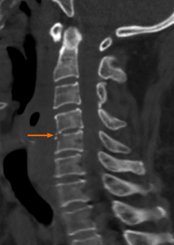
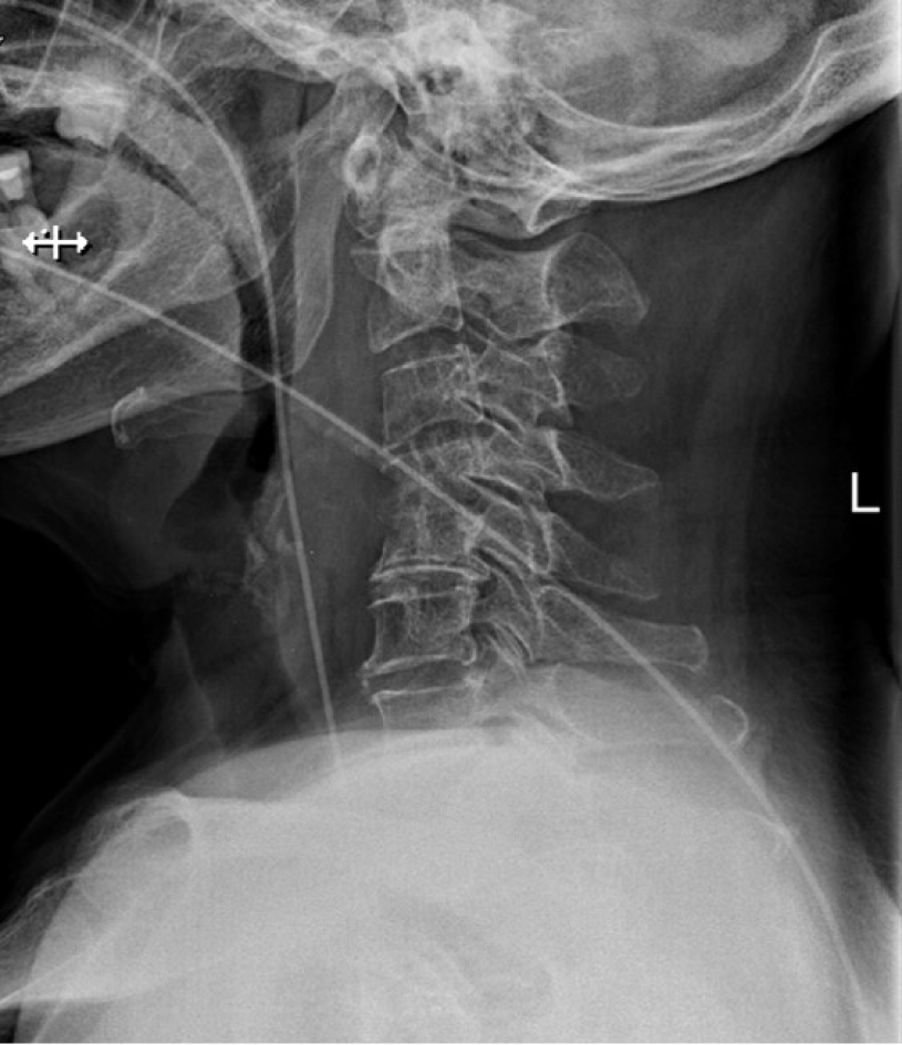
Cervical radiography and computed tomography (CT) did not find the fish bone or other foreign bodies (Figure 1). Cervical magnetic resonance imaging (MRI) demonstrated spondylitis and diskitis of C4/C5, so the patient received intravenous Biapenem 0.3 g every 8 h immediately.
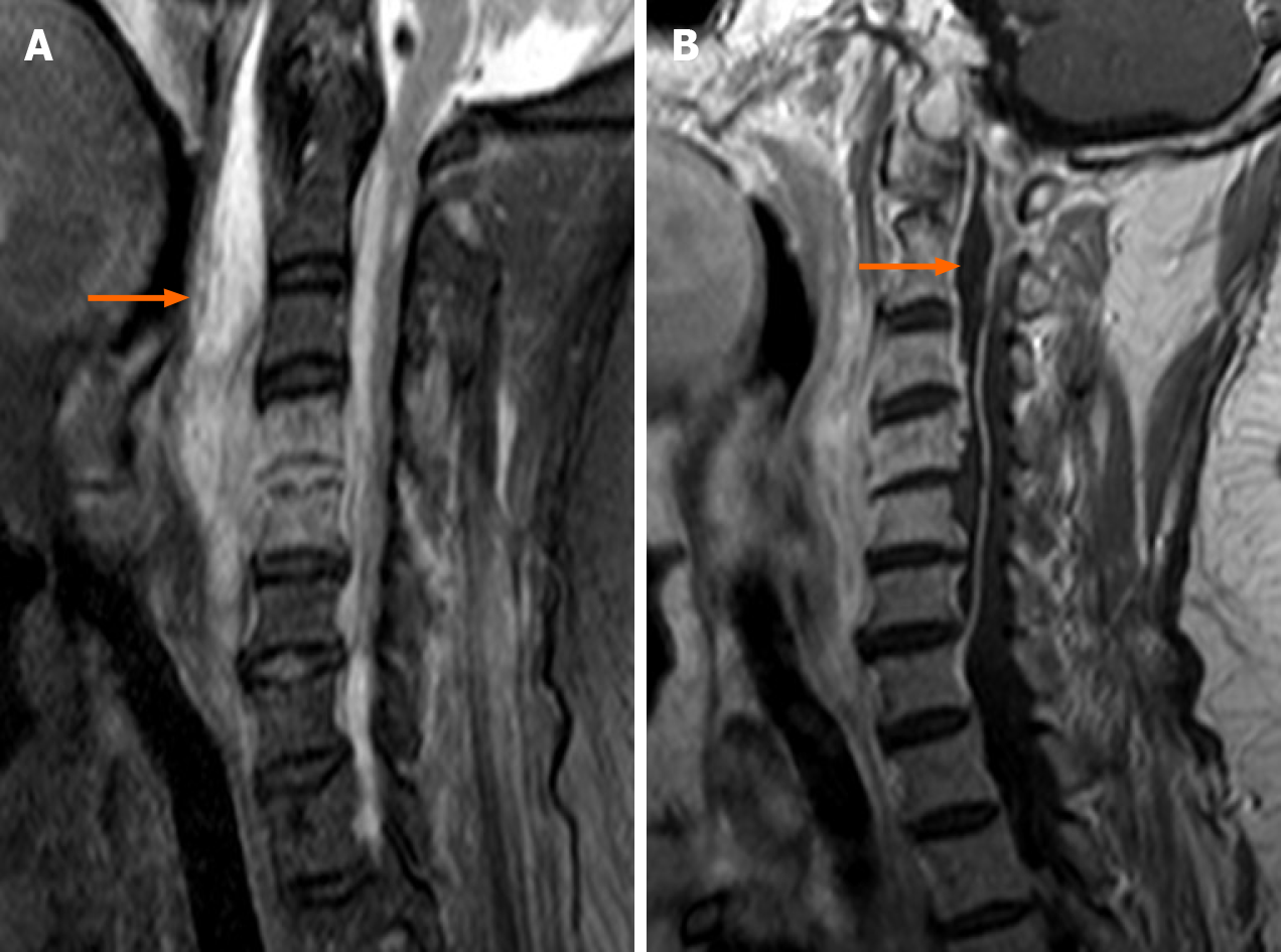
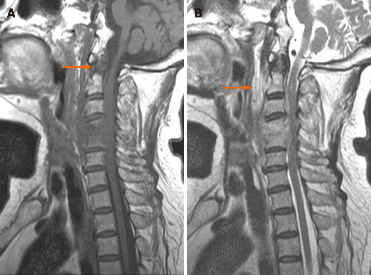
The emergency MRI revealed a great amount of septic fluid collection in the spatium retropharyngeum spreading into the spinal canal through C4/C5 disc space (Figure 2A), forming a ventral epidural abscess extending from C6 to the foramen magnum (Figure 2B).
Final diagnosis of the presented case was delayed cervical infection and incomplete quadriplegia.
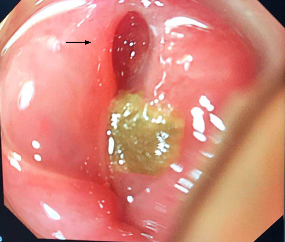
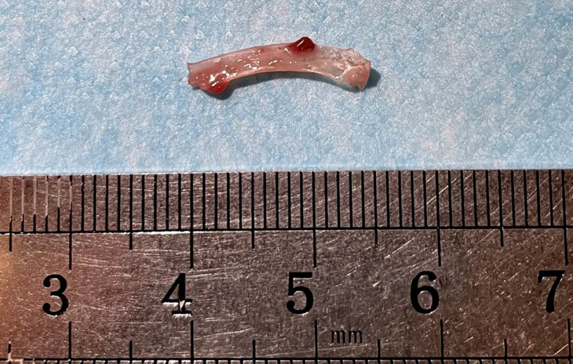
Following emergency MRI examination, emergency surgery was performed to decompress the spinal cord by removing the anterior inflammatory necrotic tissue and draining the epidural abscess. Simultaneously, flexible esophagogastroduodenoscopy found an underlying abscess and esophageal perforation, which were exposed and treated with irrigation and careful debridement (Figure 3). Enteral nutrition through an indwelling nasogastric tube was conducted, and the esophageal perforation was treated conservatively. After surgery, the fish bone was found in the irrigation fluid (Figure 4). Culture of the epidural abscess suggested Staphylococcus aureus infection.
The patient’s symptoms significantly improved postoperatively. On the second day after surgery, muscle strength of the right lower limb was improved to grade 3, and muscle strength of the other limbs was improved to grade 4. On the 15th day after surgery, MRI examination showed that the abscess in the posterior pharyngeal wall and epidural was significantly smaller than that before surgery (Figure 5). The cervical spine was stabilized with a Minerva cervicothoracic orthosis for 8 wk. Postoperative enteral nutrition through an indwelling nasogastric tube was conducted for 6 wk. Intravenous Linezolid 600 mg every 12 h was administered for 15 d until the erythrocyte sedimentation rate and CRP declined to normal levels. On the fifth week after surgery, no foreign body was found in the cervical X-ray examination (Figure 6).
One-year follow-up showed that the patient’s limb muscle strength had completely returned to normal.
Cervical epidural abscess developing from a retropharyngeal abscess and spondylitis due to upper digestive tract wall injury and perforation is extremely rare[5]. All the reported perforation cases caused by foreign body ingestion have pharyngeal or esophageal discomfort[1, 6-8]. Our patient did not complain of any symptoms, including dysphagia, odynophagia, hematemesis, dyspnea, etc. CT has a sensitivity of 90%-100% in detecting impacted fish bones[9-11], which is obviously superior to 32% by plain radiography[6]. However, in the present case, both plain radiography and CT did not identify the impacted fish bone. Physicians should be aware of the posterior pharyngeal or esophageal wall perforation in patients with cervical pyogenic spondylitis with a certain or uncertain history of foreign body ingestion, even though local discomfort symptoms are absent and the radiological examinations are negative.
Urgent management in the first 24 h is critical for the prognosis of foreign body ingestion. If the patient is subjected to delayed management for more than 2 days, the state of the local injury may develop into a fatal condition[12], especially the foreign body lodging at the esophagus entrance, which is surrounded by the cricopharyngeus muscle[13]. In the present case, symptoms due to spinal cord compression developed one month after fish bone ingestion, which missed a timely early intervention and increased the likelihood of severe complications.
Endoscopy is the most frequently used technique in clinical practice, which is recommended as the first-line therapeutic option for foreign body extraction[14]. Fish bone is one of the main ingested esophageal foreign bodies. Perforation of the esophagus caused by fish bone usually leads to mediastinitis[15-18]. In the present case, the patient developed spondylitis without mediastinitis. Endoscopy after admission revealed that the posterior wall of the laryngopharynx was ruptured and a fish bone was found in the irrigation fluid after surgery. During the diagnostic process, clinicians need to pay sufficient attention to patients with a history of fish bone ingestion, even if the immediate laryngoscopy results are negative. It is suggested that in patients without mediastinitis, esophageal perforation caused by a foreign body is mostly located in the posterior wall. In terms of treatment, we adopted local complete debridement, sufficient and effective use of antibiotics and placement of a gastric tube for nasal feeding to avoid secondary infection. These measures played a vital role in infection control and healing of the laryngopharyngeal wall perforation.
This case was our first encounter with a delayed cervical infection and incomplete quadriplegia with fish-bone ingestion and reminds us to consider the possibility of foreign body in the differential diagnosis for patients with neck pain and weakness of four limbs. It is necessary for clinicians to consider and recognize cervical infection and incomplete quadriplegia due to fish-bone ingestion in patients with atypical clinical manifestations or CT findings. We hope that the case we report here will increase awareness of cervical infection due to a foreign body and help avoid future misdiagnosis by orthopedists.
Manuscript source: Unsolicited Manuscript
Specialty type: Orthopedics
Country/Territory of origin: China
Peer-review report’s scientific quality classification
Grade A (Excellent): 0
Grade B (Very good): B
Grade C (Good): 0
Grade D (Fair): 0
Grade E (Poor): 0
P-Reviewer: Zhao N S-Editor: Ma YJ L-Editor: Webster JR P-Editor: Liu JH
| 1. | Lai AT, Chow TL, Lee DT, Kwok SP. Risk factors predicting the development of complications after foreign body ingestion. Br J Surg. 2003;90:1531-1535. [RCA] [PubMed] [DOI] [Full Text] [Cited by in Crossref: 113] [Cited by in RCA: 122] [Article Influence: 5.8] [Reference Citation Analysis (0)] |
| 2. | Wang Z, Du Z, Zhou X, Chen T, Li C. Misdiagnosis of peripheral abscess caused by duodenal foreign body: a case report and literature review. BMC Gastroenterol. 2020;20:236. [RCA] [PubMed] [DOI] [Full Text] [Full Text (PDF)] [Cited by in Crossref: 6] [Cited by in RCA: 6] [Article Influence: 1.2] [Reference Citation Analysis (0)] |
| 3. | Garrido Serrano A, Fernández Álvarez P. A foreign body in the small bowel: a rare entity of acute abdomen. Rev Esp Enferm Dig. 2019;111:84. [RCA] [PubMed] [DOI] [Full Text] [Cited by in RCA: 1] [Reference Citation Analysis (0)] |
| 4. | Costa L, Larangeiro J, Pinto Moura C, Santos M. Foreign body ingestion: rare cause of cervical abscess. Acta Med Port. 2014;27:743-748. [RCA] [PubMed] [DOI] [Full Text] [Cited by in Crossref: 8] [Cited by in RCA: 9] [Article Influence: 0.8] [Reference Citation Analysis (0)] |
| 5. | Jeon SH, Han DC, Lee SG, Park HM, Shin DJ, Lee YB. Eikenella corrodens cervical spinal epidural abscess induced by a fish bone. J Korean Med Sci. 2007;22:380-382. [RCA] [PubMed] [DOI] [Full Text] [Full Text (PDF)] [Cited by in Crossref: 11] [Cited by in RCA: 11] [Article Influence: 0.6] [Reference Citation Analysis (0)] |
| 6. | Kim HU. Oroesophageal Fish Bone Foreign Body. Clin Endosc. 2016;49:318-326. [RCA] [PubMed] [DOI] [Full Text] [Full Text (PDF)] [Cited by in Crossref: 40] [Cited by in RCA: 50] [Article Influence: 5.6] [Reference Citation Analysis (0)] |
| 7. | Peng A, Li Y, Xiao Z, Wu W. Study of clinical treatment of esophageal foreign body-induced esophageal perforation with lethal complications. Eur Arch Otorhinolaryngol. 2012;269:2027-2036. [RCA] [PubMed] [DOI] [Full Text] [Cited by in Crossref: 39] [Cited by in RCA: 48] [Article Influence: 3.7] [Reference Citation Analysis (0)] |
| 8. | Zheng SH, Li Y, Zhang SQ. Acute quadriplegia following a minimal injury in the posterior pharyngeal wall by a fishbone. CNS Neurosci Ther. 2017;23:637-639. [RCA] [PubMed] [DOI] [Full Text] [Cited by in RCA: 1] [Reference Citation Analysis (0)] |
| 9. | Liew CJ, Poh AC, Tan TY. Finding nemo: imaging findings, pitfalls, and complications of ingested fish bones in the alimentary canal. Emerg Radiol. 2013;20:311-322. [RCA] [PubMed] [DOI] [Full Text] [Cited by in Crossref: 19] [Cited by in RCA: 23] [Article Influence: 1.8] [Reference Citation Analysis (0)] |
| 10. | Jahshan F, Sela E, Layous E, Levy E, Assadi N, Shilo E, Ibrahim N, Maayan D, Ronen O. Clinical criteria for CT scan evaluation of upper digestive tract fishbone. Laryngoscope. 2018;128:2467-2472. [RCA] [PubMed] [DOI] [Full Text] [Cited by in Crossref: 9] [Cited by in RCA: 15] [Article Influence: 2.1] [Reference Citation Analysis (0)] |
| 11. | Kim EY, Min YG, Bista AB, Park KJ, Kang DK, Sun JS. Usefulness of Ultralow-Dose (Submillisievert) Chest CT Using Iterative Reconstruction for Initial Evaluation of Sharp Fish Bone Esophageal Foreign Body. AJR Am J Roentgenol. 2015;205:985-990. [RCA] [PubMed] [DOI] [Full Text] [Cited by in Crossref: 9] [Cited by in RCA: 10] [Article Influence: 1.1] [Reference Citation Analysis (0)] |
| 12. | Athanassiadi K, Gerazounis M, Metaxas E, Kalantzi N. Management of esophageal foreign bodies: a retrospective review of 400 cases. Eur J Cardiothorac Surg. 2002;21:653-656. [RCA] [PubMed] [DOI] [Full Text] [Cited by in Crossref: 100] [Cited by in RCA: 99] [Article Influence: 4.3] [Reference Citation Analysis (0)] |
| 13. | Onat S, Ulku R, Cigdem KM, Avci A, Ozcelik C. Factors affecting the outcome of surgically treated non-iatrogenic traumatic cervical esophageal perforation: 28 years experience at a single center. J Cardiothorac Surg. 2010;5:46. [RCA] [PubMed] [DOI] [Full Text] [Full Text (PDF)] [Cited by in Crossref: 34] [Cited by in RCA: 38] [Article Influence: 2.5] [Reference Citation Analysis (0)] |
| 14. | Birk M, Bauerfeind P, Deprez PH, Häfner M, Hartmann D, Hassan C, Hucl T, Lesur G, Aabakken L, Meining A. Removal of foreign bodies in the upper gastrointestinal tract in adults: European Society of Gastrointestinal Endoscopy (ESGE) Clinical Guideline. Endoscopy. 2016;48:489-496. [RCA] [PubMed] [DOI] [Full Text] [Cited by in Crossref: 274] [Cited by in RCA: 382] [Article Influence: 42.4] [Reference Citation Analysis (0)] |
| 15. | Wang J, Wu WB, Chen L, Zhu Q. Video-mediastinoscopy assisted fish bone extraction and superior Medistinal abscess debridement. J Cardiothorac Surg. 2018;13:38. [RCA] [PubMed] [DOI] [Full Text] [Full Text (PDF)] [Cited by in Crossref: 6] [Cited by in RCA: 8] [Article Influence: 1.1] [Reference Citation Analysis (0)] |
| 16. | Iida T, Nakagaki S, Satoh S, Shimizu H, Kaneto H. Image of the month: Severe Mediastinitis and Pneumomediastinum Following Esophageal Perforation by a Fish Bone. Am J Gastroenterol. 2015;110:1262. [RCA] [PubMed] [DOI] [Full Text] [Cited by in RCA: 1] [Reference Citation Analysis (0)] |
| 17. | Mariani AW, Pêgo-Fernandes PM, Samano MN, de Albuquerque Cavalcanti EF, Cevasco JJ, Gattaz MD. Indolent form of mediastinitis caused by oesophageal perforation from fish bone ingestion. BMJ Case Rep. 2009;2009. [RCA] [PubMed] [DOI] [Full Text] [Cited by in Crossref: 1] [Cited by in RCA: 3] [Article Influence: 0.2] [Reference Citation Analysis (0)] |
| 18. | An JS, Baek IH, Chun SY, Kim KO. Successful endoscopic band ligation of esophageal perforation by fish bone ingestion. J Laparoendosc Adv Surg Tech A. 2013;23:459-462. [RCA] [PubMed] [DOI] [Full Text] [Cited by in Crossref: 2] [Cited by in RCA: 3] [Article Influence: 0.3] [Reference Citation Analysis (0)] |


