Published online Sep 6, 2021. doi: 10.12998/wjcc.v9.i25.7478
Peer-review started: January 5, 2021
First decision: May 11, 2021
Revised: May 23, 2021
Accepted: July 16, 2021
Article in press: July 16, 2021
Published online: September 6, 2021
Processing time: 237 Days and 20.3 Hours
Forearm crisscross injury is rare in children; there is no relevant literature so far. Surgeons lack experience and knowledge in treating this type of crisscross injury. We report a case of forearm crisscross injury in a child for the first time and analyze its mechanism.
An 8-year-old boy experienced pain in his left forearm when he accidentally fell while skateboarding. Physical examination revealed swelling and deformity of the left forearm. We performed imaging and the results revealed left radial head dislocation, left distal radial epiphyseal separation from the shaft, and interruption of the continuity of the dorsal cortex of the left distal ulna. Anteroposterior and lateral X-ray films showed that the radius and ulna were criss
This is the first report of a child with a forearm crisscross injury in which the mechanism and the differences from adult crisscross injury are analyzed. Minimally invasive surgery with intramedullary fixation can achieve a good therapeutic effect. This case provides a reference for the treatment of similar patients in the future.
Core Tip: Forearm crisscross injury is rare in children; there is no relevant literature so far. This case highlights the first pediatric case of a forearm crisscross injury and analyze its differences in the mechanism from injury in adult patients, which could improve the understanding of forearm crisscross injury in children.
- Citation: Jiang YK, Wang YB, Peng CG, Qu J, Wu DK. First case of forearm crisscross injury in children: A case report. World J Clin Cases 2021; 9(25): 7478-7483
- URL: https://www.wjgnet.com/2307-8960/full/v9/i25/7478.htm
- DOI: https://dx.doi.org/10.12998/wjcc.v9.i25.7478
Forearm crisscross injury is rare due its special mechanism; less than ten cases have been reported in the existing literature. Among current reports, all forearm crisscross injuries occurred in adult patients, and there have been no reports on pediatric patients. We report the first pediatric case of a forearm crisscross injury and analyze its differences in the mechanism from injury in adult patients, which could improve the understanding of forearm crisscross injury in children. Minimally invasive surgery led to the patient’s smooth recovery at follow-up week 14. This case provided a reference for diagnosis and treatment of forearm crisscross injuries in children.
An 8-year-old patient experienced pain and deformity in his left forearm due to an accidental fall. He was admitted to our hospital 2 h after semi-restriction of movement.
A detailed medical history inquiry revealed that the patient accidentally fell while skateboarding; his palm touched the ground, causing excessive external rotation of his forearm and resulting in injury.
Physical examination revealed ecchymosis, deformity of the left wrist, and limited range of motion due to pain. The left forearm showed no obvious swelling, and there was no paresthesia of the left upper extremity, and no vascular or nerve injuries.
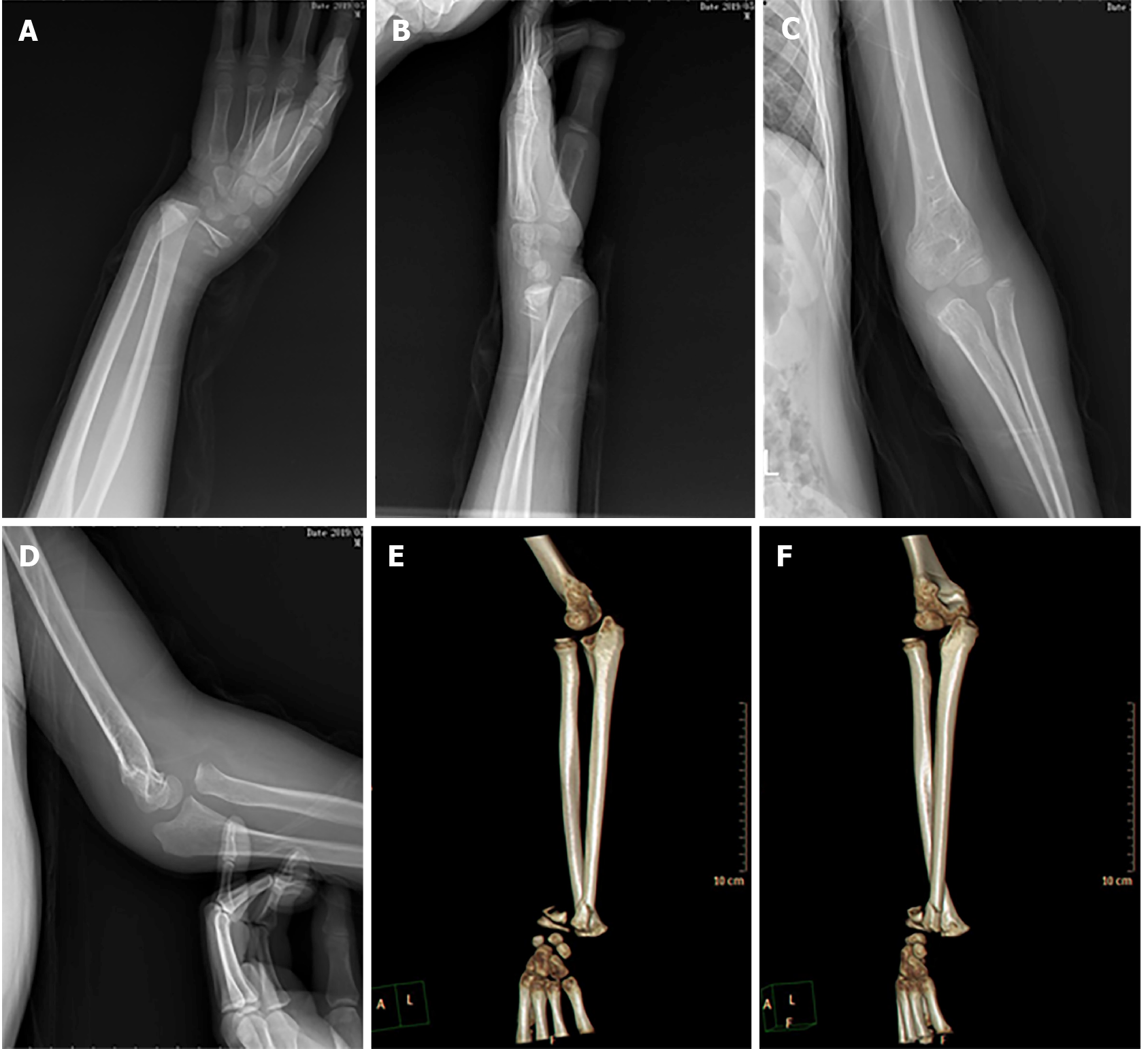
We performed X-ray and 3-dimensional computed tomography, which showed that the left elbow humeroradial joint was poorly aligned and the radial head was dislocated forward (Figure 1). The left distal radial epiphysis was separated from the shaft, indicating a Salter-Harris type II epiphyseal injury. The cortical continuity of the left distal ulna was interrupted. Anterior and lateral radiographs showed that the ulna and radius were crisscrossed.
A diagnosis of superior radioulnar joint dislocation, left distal radial epiphyseal injury, and left distal ulnar fracture was made.
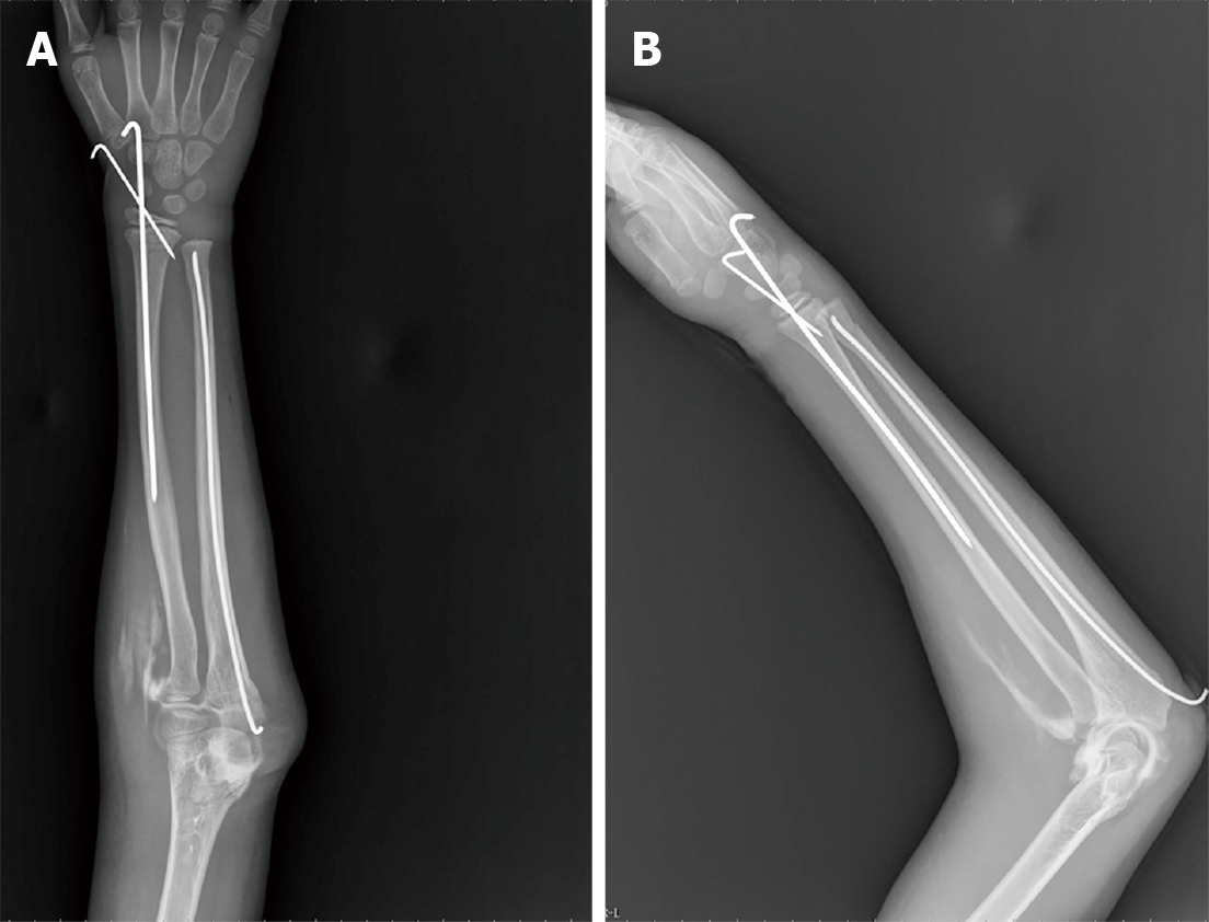
None of the examinations suggested any obvious contraindications; hence, manual reduction was performed under anesthesia. Unfortunately, manual reduction failed and open reduction surgery was planned. After anesthesia induction, a 5-cm longitudinal incision was made on the radial side of the palmar side of the left wrist to expose the distal radius. The anterior spiral muscle was embedded in the distal radius fracture. The embedded soft tissue was removed and the distal radius fracture was fully reduced under direct vision. Crossed K-wire fixation was performed. When the distal ulnar and radial anatomical structures were restored, the original anatomical position of the superior radioulnar joint was also automatically restored. Afterward, the distal ulnar fracture was fixed using a 2.0-mm elastic intramedullary needle.
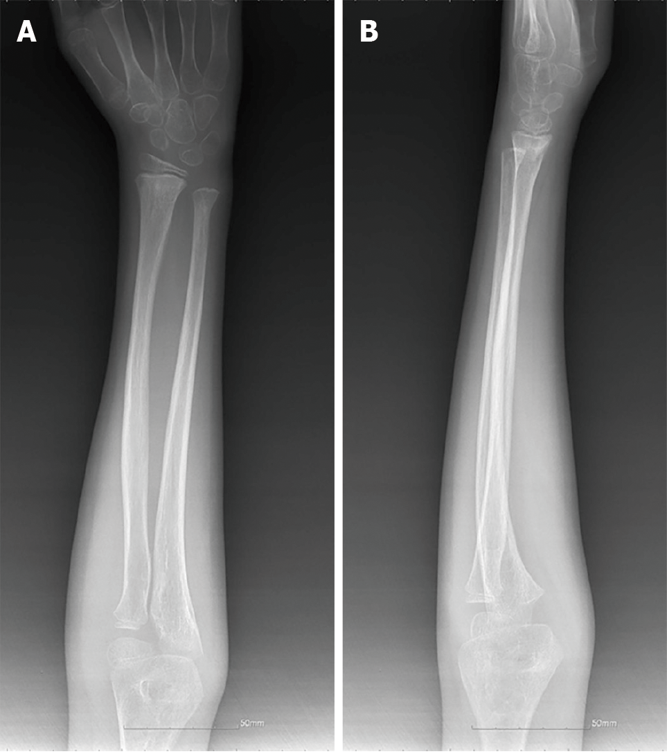
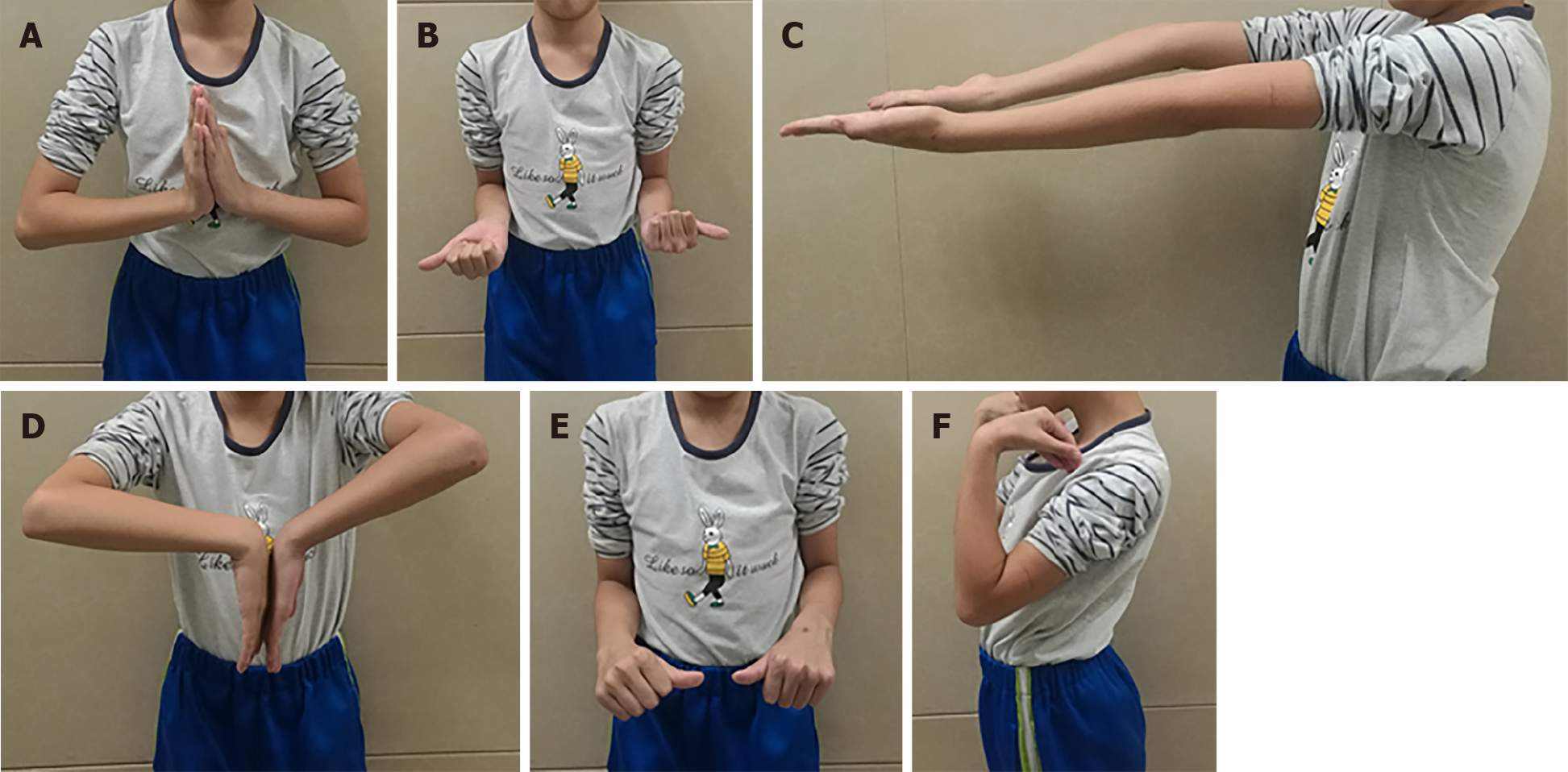
Postoperatively, the left elbow was flexed to 90° and the forearm was in a neutral position (Figure 2). After 6 wk of plaster cast immobilization, X-ray examination showed that the fracture fully healed and the disabilities of the arm, shoulder, and hand (DASH) score was 16 points. Hence, the plaster and internal fixation were removed and functional exercises were started (Figure 3). At follow-up week 14, the patient had good left upper extremity function without complaints of pain or limited range of motion (Figure 4). The DASH upper extremity function score was 9 points, with good follow-up results.
Forearm crisscross injury is rare. Due to the special mechanism, less than ten cases have been reported worldwide; all cases involved adult patients[1,2]. To date, this special injury has not been reported in children; its pathogenesis, treatment, and prognosis remain unclear. Leung et al[2] first reported the definition and diagnostic criteria of crisscross injury in 2002. A currently recognized crisscross injury refers to simultaneous upper and lower radioulnar joint dislocation with intact interstitial membrane, accompanied by fractures of the radial head and the ulnar styloid process, but not accompanied by ipsilateral ulnar and radial shaft fractures. Lateral and anteroposterior radiographic findings of the forearm revealed radioulnar crisscross. According to Leung et al[2], crisscross injuries can be classified into two types: Type I refers to forward radial and ulnar head dislocations; type II refers to posterior dislocation of the radial and ulnar heads. The former is due to forearm overpronation, while the latter is caused by supination (with the intact interosseous membrane serving as a fulcrum that participates in the mechanism of the forearm crisscross injury)[3]. Our patient’s imaging data showed that the ulna and radius were crisscrossed in the anterior and lateral X-ray images. Although we did not perform magnetic resonance imaging to prove that the interosseous membrane of the forearm was intact, the patient's forearm was not swollen. When the anatomical structure of the distal ulna and radius recovered, the superior radioulnar joint was automatically restored without any additional intervention. These results indirectly proved that the interosseous membrane was intact. In our case, strictly speaking, only the superior radioulnar joint was dislocated. The distal radius epiphysis was fractured and some distal joints were still connected to the ulna; hence, the inferior radioulnar joint did not comprise a true dislocation. Since the ligament strength in children is greater than that of the epiphyseal plate, epiphyseal injury occurs in the distal radius without inferior radioulnar separation. We believe that this is a special manifestation of crisscross injury in children.
In the diagnosis of crisscross injury in adults, simultaneous dislocation of the superior and inferior radioulnar joints without combined ipsilateral ulnar and radial shaft fractures is a universally accepted diagnostic criterion. Current reports include no description of fractures other than the radioulnar joint dislocation[2,4,5]. During adolescence, tendons, ligaments, and joint capsules are two to five times stronger than epiphyses plates[6]. Therefore, when radioulnar joint dislocations occur in children, their forearms are extremely pronated or supinated, and injuries to the epiphyseal plate” are possible. In our case, the patient's forearm was extremely externally rotated and the shearing force of the epiphysis and ligament of the distal radius increased. Epiphyseal fracture occurred due to the different strengths of the epiphyses and ligaments. The child’s single ulna was unable to support the weight of the body and fracture. Our case was different from those reported in the existing literature, possibly due to the differences in the strength of bones and ligaments of children from those of adults. Our patient’s imaging manifestation is fully consistent with the diagnosis of a type I crisscross injury, ignoring the impact of distal radial epiphyseal fractures and considering the palm and ulna as a whole. For fracture in children, we generally do not perform CT examination before operation. However, this is a rare and unique case. In order to observe the relative position of the ulna and radius after injury, we chose three-dimensional CT examination. In the process of CT examination, we have made full radiation protection to the non-examined parts of the children, so the amount of radiation received by the children is not much.
Cases of crisscross injuries reported in the existing literature have been successfully treated by closed reduction, manipulation, plaster, or external fixation, and no complications of joint instability or pain occurred. In a patient reported by Potter et al[4], closed reduction failed multiple times due to deformities of the radial head, and scar reduction was ultimately successful. In our case, multiple manual reductions failed and the patient had an open epiphyseal fracture. Therefore, epiphyseal fractures must be properly treated as early as possible, focusing on anatomical reduction; otherwise, late deformities can easily occur due to premature epiphyseal closure. We performed intramedullary fixation to treat the fracture, reducing the patient’s traumatic stress, while achieving anatomical reduction. The patient fully recovered and returned to his normal life 3 mo postoperatively.
We report a case of forearm crisscross injury in children for the first time and analyze the mechanism and differences from adults. Minimally invasive surgery with intramedullary fixation for a forearm crisscross fracture can achieve good results. This case provides a reference for future diagnosis and treatment of similar patients.
Manuscript source: Unsolicited manuscript
Specialty type: Orthopedics
Country/Territory of origin: China
Peer-review report’s scientific quality classification
Grade A (Excellent): 0
Grade B (Very good): B
Grade C (Good): C
Grade D (Fair): 0
Grade E (Poor): 0
P-Reviewer: Bianchi F, Pop TL S-Editor: Liu M L-Editor: Wang TQ P-Editor: Ma YJ
| 1. | Papageorgiou C, Vradelis K. Simultaneous dislocation of the proximal and distal radioulnar joints. A case report. Acta Orthop Scand. 1995;66:180. [RCA] [PubMed] [DOI] [Full Text] [Cited by in Crossref: 9] [Cited by in RCA: 9] [Article Influence: 0.3] [Reference Citation Analysis (0)] |
| 2. | Leung YF, Ip SP, Wong A, Wong KN, Wai YL. Isolated dislocation of the radial head, with simultaneous dislocation of proximal and distal radio-ulnar joints without fracture in an adult patient: case report and review of the literature. Injury. 2002;33:271-273. [RCA] [PubMed] [DOI] [Full Text] [Cited by in Crossref: 14] [Cited by in RCA: 16] [Article Influence: 0.7] [Reference Citation Analysis (0)] |
| 3. | Wong BL, Rama KB, Kumar A. Simultaneous dislocation of proximal and distal radio-ulnar joints: a case report. J Orthop Surg (Hong Kong). 2013;21:106-109. [RCA] [PubMed] [DOI] [Full Text] [Cited by in Crossref: 4] [Cited by in RCA: 5] [Article Influence: 0.4] [Reference Citation Analysis (0)] |
| 4. | Potter M, Wang A. Simultaneous dislocation of the radiocapitellar and distal radioulnar joints without fracture: case report. J Hand Surg Am. 2012;37:2502-2505. [RCA] [PubMed] [DOI] [Full Text] [Cited by in Crossref: 6] [Cited by in RCA: 7] [Article Influence: 0.5] [Reference Citation Analysis (0)] |
| 5. | Falsafi M, Baghianimoghadam B. Simultaneous Unilateral Radial Shaft Fracture With Distal and Proximal Radioulnar Joint Dislocation, An Extremely Rare Case Report. Rev Med Chir Soc Med Nat Iasi. 2016;120:619-622. [PubMed] |
| 6. | Peiró A, Aracil J, Martos F, Mur T. Fractures of the distal tibial epiphysis in adolescence. J Bone Joint Surg Am. 1983;65:1208. [PubMed] |


