Published online Jan 16, 2021. doi: 10.12998/wjcc.v9.i2.416
Peer-review started: August 6, 2020
First decision: November 8, 2020
Revised: November 18, 2020
Accepted: November 29, 2020
Article in press: November 29, 2020
Published online: January 16, 2021
Processing time: 154 Days and 5.8 Hours
Fecal impaction is defined as a large mass of compacted feces in the colon and has the potential to induce a serious medical condition in elderly individuals. Fecal impaction is generally preventable, and early recognition of the typical radiological findings is important for making an early diagnosis. The factors that lead to fecal impaction are usually similar to those causing constipation. Few cases with fecal impaction associated with a diverticulum have been reported.
We present the case of a 62-year-old woman who suffered from abdominal pain and vomiting, had a medical history of repeated acute abdomen and was diagnosed with fecal impaction in the descending colon based on X-ray and computed tomography (CT) imaging. After examination by gastrografin-enhanced colonography following colonoscopy and CT colonography, the fecalith was suspected to have been produced at the site of a large diverticulum in the transverse colon. The fecalith was surgically resected, and a histological diagnosis of pseudodiverticulum was made. There was no recurrence during 33 mo of follow-up.
This case highlights the importance of accurate identification and treatment of a fecal impaction. This case indicated that the endoscopic evacuation and subsequent colonography were effective for identifying a diverticulum that might have caused fecal impaction. A fecal impaction was associated with the diverticulum. Consequently, the planned diverticulectomy was performed. Appropriate emergency medical treatment and maintenance treatments should be selected in such cases to prevent recurrence.
Core Tip: A 62-year-old woman presented to the emergency department with abdominal pain and vomiting. She had several medical histories of treatment for ileus over the past four years, but no specific findings were detected by colonoscopy. Fecal impaction was observed by computed tomography (CT), and the fecalith was broken and removed by colonoscopy. CT colonography verified the presence of a colonic diverticulum, which was suspected to have been responsible for the fecalith. After the surgery on the diverticulum, the patient became free from any further episodes of abdominal pain. A fecalith can be caused by a giant diverticulum that is not evident on colonoscopy.
- Citation: Tanabe H, Tanaka K, Goto M, Sato T, Sato K, Fujiya M, Okumura T. Rare case of fecal impaction caused by a fecalith originating in a large colonic diverticulum: A case report. World J Clin Cases 2021; 9(2): 416-421
- URL: https://www.wjgnet.com/2307-8960/full/v9/i2/416.htm
- DOI: https://dx.doi.org/10.12998/wjcc.v9.i2.416
Fecal impaction is a medical disorder that is defined as a large mass of compacted feces that cannot be spontaneously evacuated. This condition sometimes induces life-threatening complications such as stercoral colitis, which is caused by increased intraluminal pressure[1,2]. Patients with fecal impaction should receive immediate treatment, including manual disimpaction or endoscopic fragmentation[3]. The factors leading to fecal impaction are similar to those causing chronic constipation. These factors are associated with lifestyle, anatomic, mechanical, neurologic, metabolic, and medication-related conditions. Institutionalized or hospitalized elderly patients and patients with neuropsychiatric disease are at high risk for developing fecal impaction[4]. The management of fecal impaction is divided into three main components: Disimpaction, evacuation of the colon, and administration of a bowel maintenance regimen. Maintenance treatments should be conducted to prevent recurrence. Polyethylene glycol plus electrolytes has recently been used as a maintenance regimen. We herein report a very rare case in which recurrent fecal impaction was caused by a large diverticulum. The patient was successfully treated with colonoscopic disimpaction and diverticulectomy.
A 62-year-old woman presented to the emergency department with abdominal pain and vomiting.
Her medical history included hospitalization for treatment for ileus and ischemic colitis several times in the past four years; however, no specific findings were detected by colonoscopy at those times.
The patient had an unremarkable past illness.
The patient has no specific family history.
On evaluation in our emergency department, no abdominal mass or rebound tenderness was found in a physical examination.
The initial laboratory examinations revealed an increased white blood cell count (11.8 × 10³/mm3), normal red blood cell count (4.27 × 10⁶/mm3), normal platelet count (237 × 103/mm3), and a normal C-reactive protein level (0.05 mg/dL), indicating acute inflammation.
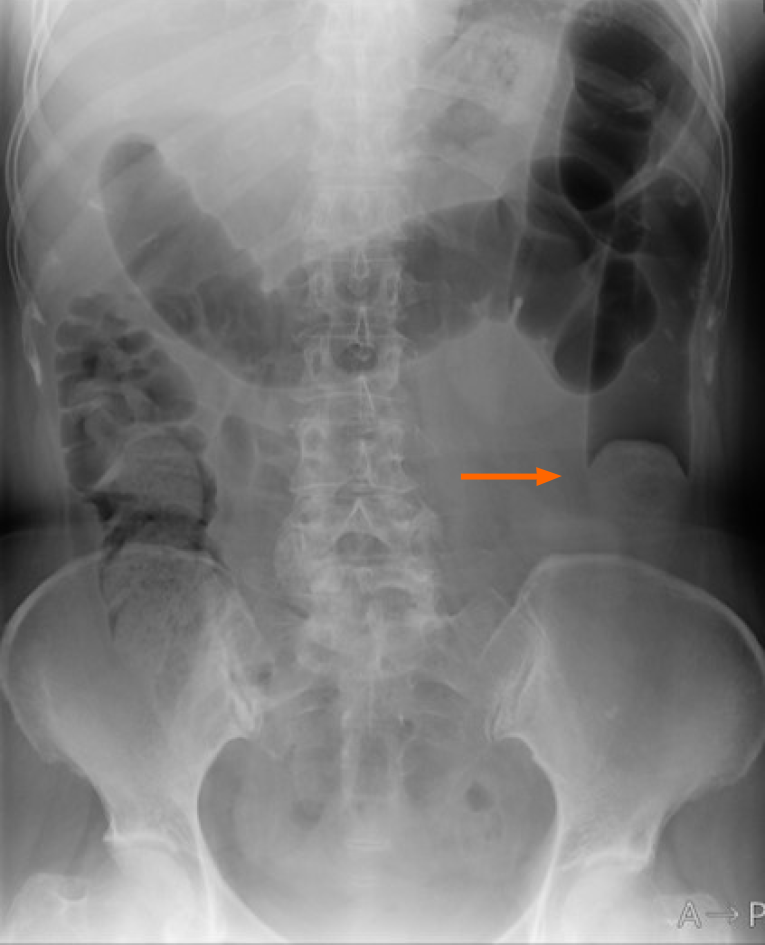
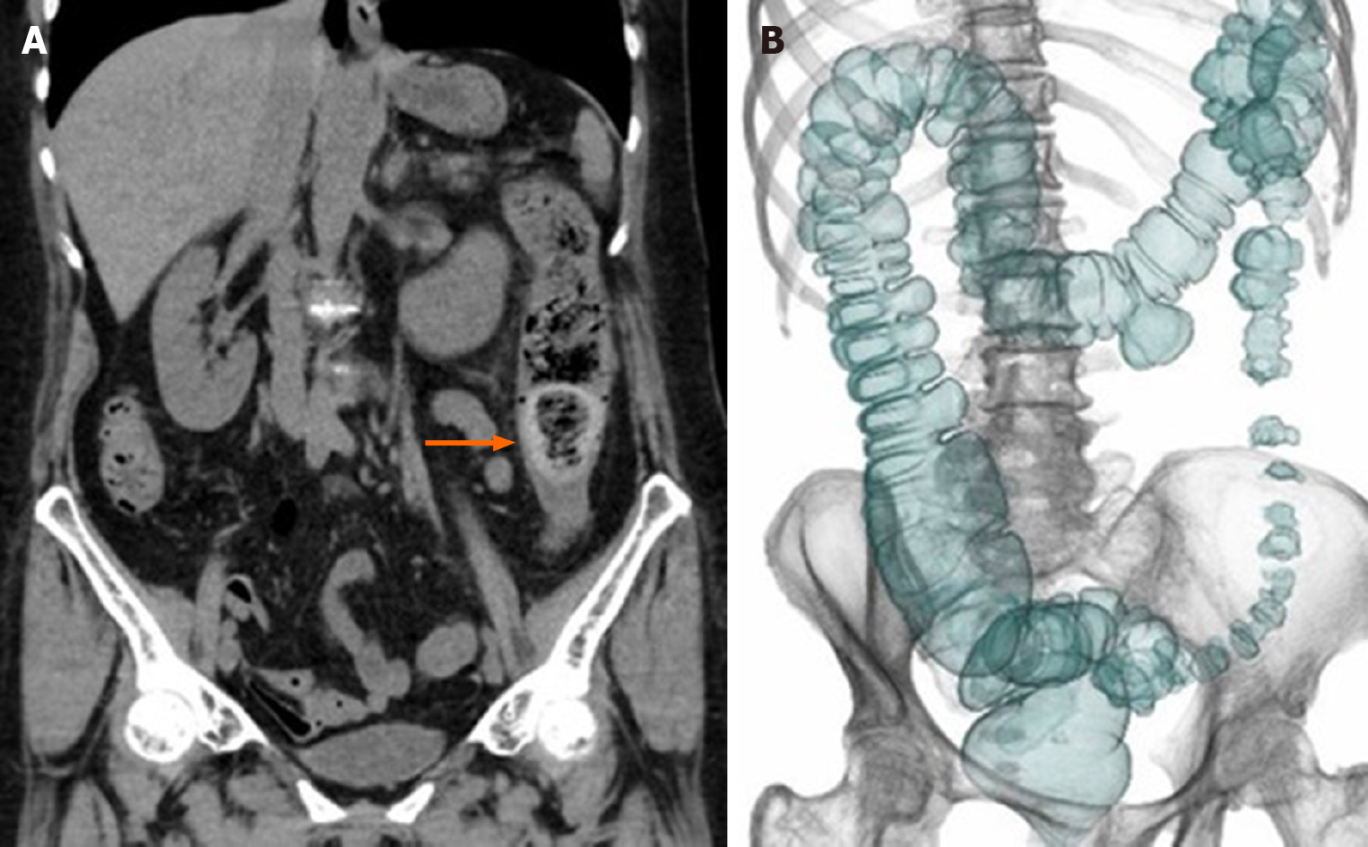
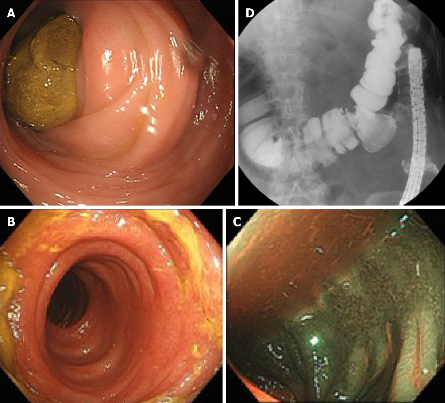
An X-ray film revealed colonic gas filling from the cecum to the descending colon with a defect near the sigmoid-descending junction (Figure 1). Abdominal computed tomography (CT) revealed intracolonic feces and fecal impaction in the descending colon (Figure 2A). On the following day, colonoscopy was performed without preparation and an impacted fecaloma was found in the descending colon (Figure 3A). It was crushed with jumbo forceps and removed by water-jet injection. Inflammatory findings, including redness and swelling on the oral side of the impacted lesion, were observed; the appearance was similar to ischemic colitis (Figure 3B and C). Gastrografin-contrast colonography revealed a diverticulum 35 mm in diameter in the transverse colon (Figure 3D). After obtaining relief from her symptoms, CT colonography revealed stenosis in the descending colon and a transverse colonic diverticulum, which was suspected to have been responsible for the fecalith (Figure 2B).
The final diagnosis of the presented case was acute abdomen with fecal impaction caused by a fecalith originating in a large colonic diverticulum.
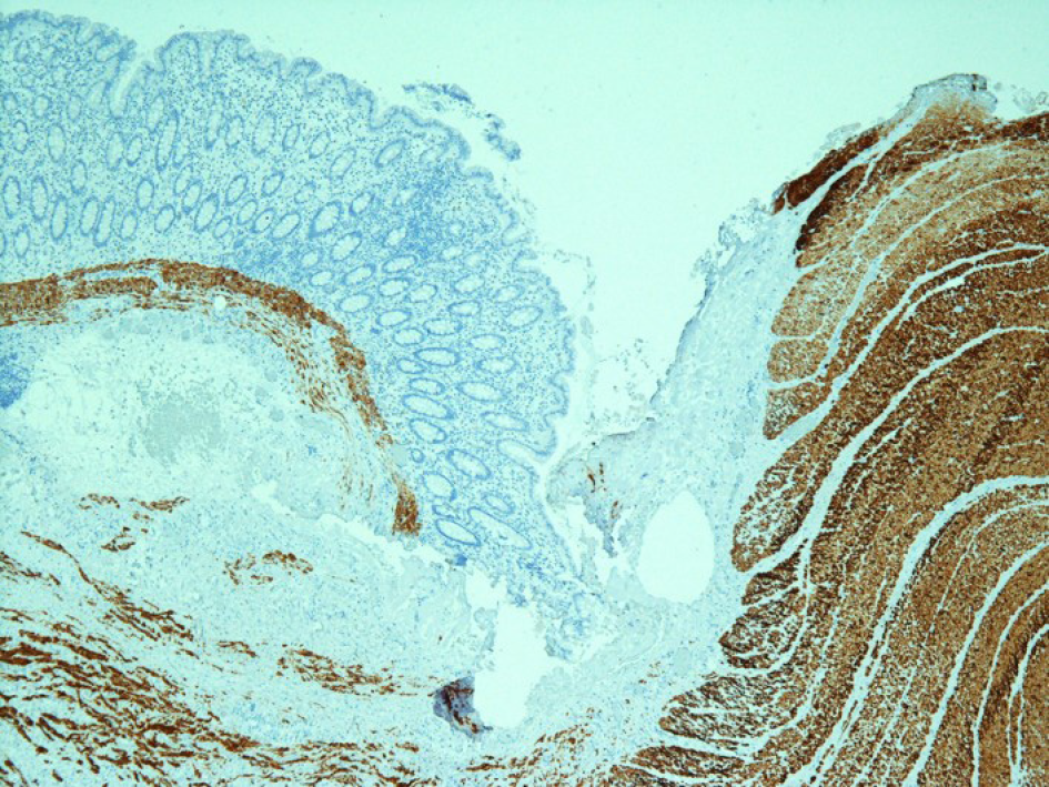
The large diverticulum was surgically resected. A histological examination with desmin immunostaining revealed a pseudodiverticulum without a muscularis propria (Figure 4).
The patient was prescribed magnesium oxide. The patient did not experience any further episodes of abdominal pain during 33 mo of follow-up.
The diverticulum observed in this case was 35 mm in diameter, indicating a large diverticulum. Mechanical and/or functional disorders were suspected based on her history of hospitalization. The diverticulum was not detected with CT at either of her previous hospitalizations. Colonoscopy suggested possible ischemic colitis. Neither examination indicated the existence of a large diverticulum because the size was equivalent to the diameter of the colon and the internal appearance was similar to the surface of the normal colonic mucosa. Fortunately, no severe complications arose due to the diverticulum in the transverse colon.
A systematic review reported that the symptoms that accompany diverticula include — but are not limited to — abdominal pain, constipation, diarrhea, vomiting, and nausea. Since these symptoms are not specific for the manifestation, radiological imaging is very important[5]. CT is thought to be the most accurate imaging modality and is recommended for decision-making in relation to treatment. Giant colonic diverticula can become complicated due to perforation, infection, obstruction, bleeding or fistulas[6]. After primary emergency medical treatments, diverticulectomy is proposed as a curable treatment. With recent advances in radiological imaging, CT colonography has become a noninvasive test for colorectal disorders[7]. It can accurately detect the presence of diverticula and provide important clinical information. Clinical guidelines mention that diverticulosis is more commonly demonstrated on CT colonography than by colonoscopy and that colonoscopy is more sensitive for the detection of colitis[8]. At the first medical examination in our emergency department, a remarkable radiological finding of fecal impaction was observed on an X-ray film, which showed a concave air image. CT showed an impacted fecalith in the descending colon but not the diverticulum in the transverse colon. The primary treatment for impaction was endoscopic evacuation and concomitant gastrografin-contrast colonography was conducted to indicate the diverticulum[9]. CT colonoscopy clearly presented the diverticulum and stenotic colon, demonstrating fecal impaction from the diverticulum. Our detailed radiological examinations with CT colonoscopy and colonography might have yielded an accurate diagnosis of diverticulum that was previously overlooked on standard colonoscopy[10]. Consequently, surgical treatment was conducted and a pseudodiverticulum was histologically diagnosed. Laparoscopic diverticulectomy has been recently reported as less invasive[11]. This less invasive treatment should have been planned in our case.
Cases of transverse colon diverticulitis with a fecalith have only rarely been reported[12]. Furthermore, there is no previously published study showing a fecalith that had originated in a transverse colonic diverticulum and had caused impaction in the distal colon, as far as we could determine based on a database search of PubMed using the following terms: Diverticulum and (fecalith or “stercoral colitis”). Meckel’s diverticulum is associated with clinical conditions similar to those stemming from a fecalith in the small intestine. Meckel’s diverticulum is a congenital anomaly of the gastrointestinal tract that is found in 2% of the young population[13,14]. Enteroliths are found in approximately 0.2%-10% of patients with Meckel’s diverticulum. Extrusion of an enterolith into the intestinal lumen causes small bowel obstruction. Surgical treatment involves en bloc resection of the impacted stone and diverticulum. Similarly in our case, surgical treatment successfully eliminated the repeated symptoms that had been occurring in the patient.
We herein report a very rare case in which fecal impaction prolapsed from a large diverticulum. It was important to precisely diagnose the fecalith and to remove it presently by colonoscopy. Diverticulectomy prevented recurrence until the time of writing this report. This case highlights the importance of accurate identification and treatment of a fecal impaction.
Manuscript source: Unsolicited manuscript
Specialty type: Medicine, research and experimental
Country/Territory of origin: Japan
Peer-review report’s scientific quality classification
Grade A (Excellent): 0
Grade B (Very good): B, B
Grade C (Good): 0
Grade D (Fair): D
Grade E (Poor): 0
P-Reviewer: Huo Q, Yao D S-Editor: Gao CC L-Editor: A P-Editor: Liu JH
| 1. | Hussain ZH, Whitehead DA, Lacy BE. Fecal impaction. Curr Gastroenterol Rep. 2014;16:404. [RCA] [PubMed] [DOI] [Full Text] [Cited by in Crossref: 34] [Cited by in RCA: 30] [Article Influence: 3.0] [Reference Citation Analysis (0)] |
| 2. | Serrano Falcón B, Barceló López M, Mateos Muñoz B, Álvarez Sánchez A, Rey E. Fecal impaction: a systematic review of its medical complications. BMC Geriatr. 2016;16:4. [RCA] [PubMed] [DOI] [Full Text] [Full Text (PDF)] [Cited by in Crossref: 92] [Cited by in RCA: 71] [Article Influence: 7.9] [Reference Citation Analysis (0)] |
| 3. | Corban C, Sommers T, Sengupta N, Jones M, Cheng V, Friedlander E, Bollom A, Lembo A. Fecal Impaction in the Emergency Department: An Analysis of Frequency and Associated Charges in 2011. J Clin Gastroenterol. 2016;50:572-577. [RCA] [PubMed] [DOI] [Full Text] [Cited by in Crossref: 19] [Cited by in RCA: 21] [Article Influence: 2.3] [Reference Citation Analysis (2)] |
| 4. | Sommers T, Petersen T, Singh P, Rangan V, Hirsch W, Katon J, Ballou S, Cheng V, Friedlander D, Nee J, Lembo A, Iturrino J. Significant Morbidity and Mortality Associated with Fecal Impaction in Patients Who Present to the Emergency Department. Dig Dis Sci. 2019;64:1320-1327. [RCA] [PubMed] [DOI] [Full Text] [Cited by in Crossref: 15] [Cited by in RCA: 13] [Article Influence: 2.2] [Reference Citation Analysis (0)] |
| 5. | Ünal E, Onur MR, Balcı S, Görmez A, Akpınar E, Böge M. Stercoral colitis: diagnostic value of CT findings. Diagn Interv Radiol. 2017;23:5-9. [RCA] [PubMed] [DOI] [Full Text] [Cited by in Crossref: 16] [Cited by in RCA: 24] [Article Influence: 3.0] [Reference Citation Analysis (0)] |
| 6. | Nigri G, Petrucciani N, Giannini G, Aurello P, Magistri P, Gasparrini M, Ramacciato G. Giant colonic diverticulum: clinical presentation, diagnosis and treatment: systematic review of 166 cases. World J Gastroenterol. 2015;21:360-368. [RCA] [PubMed] [DOI] [Full Text] [Full Text (PDF)] [Cited by in CrossRef: 56] [Cited by in RCA: 48] [Article Influence: 4.8] [Reference Citation Analysis (35)] |
| 7. | De Cecco CN, Ciolina M, Annibale B, Rengo M, Bellini D, Muscogiuri G, Maruotti A, Saba L, Iafrate F, Laghi A. Prevalence and distribution of colonic diverticula assessed with CT colonography (CTC). Eur Radiol. 2016;26:639-645. [RCA] [PubMed] [DOI] [Full Text] [Cited by in Crossref: 32] [Cited by in RCA: 28] [Article Influence: 3.1] [Reference Citation Analysis (0)] |
| 8. | Nagata N, Ishii N, Manabe N, Tomizawa K, Urita Y, Funabiki T, Fujimori S, Kaise M. Guidelines for Colonic Diverticular Bleeding and Colonic Diverticulitis: Japan Gastroenterological Association. Digestion. 2019;99 Suppl 1:1-26. [RCA] [PubMed] [DOI] [Full Text] [Cited by in Crossref: 76] [Cited by in RCA: 128] [Article Influence: 21.3] [Reference Citation Analysis (0)] |
| 9. | Gu L, Ding C, Tian H, Yang B, Zhang X, Hua Y, Gong J, Li N. Use of gastrografin in the management of faecal impaction in patients with severe chronic constipation: a randomized clinical trial. ANZ J Surg. 2019;89:239-243. [RCA] [PubMed] [DOI] [Full Text] [Cited by in Crossref: 4] [Cited by in RCA: 6] [Article Influence: 0.9] [Reference Citation Analysis (0)] |
| 10. | Bernal-Sprekelsen JC, Valderas Cortés GF, Gómez Romero L. Fecal incontinence in older patients: A narrative review. Cir Esp. 2018;96:391-392. [RCA] [PubMed] [DOI] [Full Text] [Cited by in Crossref: 1] [Cited by in RCA: 1] [Article Influence: 0.1] [Reference Citation Analysis (0)] |
| 11. | Benlice C, Costedio M, Stocchi L, Abbas MA, Gorgun E. Hand-assisted laparoscopic vs open colectomy: an assessment from the American College of Surgeons National Surgical Quality Improvement Program procedure-targeted cohort. Am J Surg. 2016;212:808-813. [RCA] [PubMed] [DOI] [Full Text] [Cited by in Crossref: 16] [Cited by in RCA: 12] [Article Influence: 1.3] [Reference Citation Analysis (0)] |
| 12. | Solak A, Solak I, Genç B, Sahin N, Yalaz S. Transverse colon diverticulitis with calcified fecalith. Eurasian J Med. 2013;45:68-70. [RCA] [PubMed] [DOI] [Full Text] [Cited by in Crossref: 3] [Cited by in RCA: 3] [Article Influence: 0.3] [Reference Citation Analysis (0)] |
| 13. | Lai HC. Intestinal obstruction due to Meckel's enterolith. Pediatr Neonatol. 2010;51:139-140. [RCA] [PubMed] [DOI] [Full Text] [Cited by in Crossref: 10] [Cited by in RCA: 10] [Article Influence: 0.7] [Reference Citation Analysis (0)] |
| 14. | Field RJ Sr, Field RJ Jr. Intestinal obstruction produced by fecalith arising in Meckel's diverticulum. AMA Arch Surg. 1959;79:8-9. [RCA] [PubMed] [DOI] [Full Text] [Cited by in Crossref: 5] [Cited by in RCA: 7] [Article Influence: 0.3] [Reference Citation Analysis (0)] |


